File:Male XY karyotype.jpg
Male_XY_karyotype.jpg (311 × 400 pixels, file size: 5 KB, MIME type: image/jpeg)
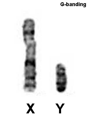
Male XY karyotype
X and Y karyotype with G-banding labelling. Note the difference in overall size of the 2 sex chromosomes.
G-banding - chromosome banding pattern seen by trypsin treatment and then staining with the dye Giemsa stain.
Metaphase is a cell division term referring to the third mitotic stage, mitotic spindle kinetochore microtubules align chromosomes in one midpoint plane. Metaphase ends when sister kinetochores separate. Originally based on light microscopy of living cells and electron microscopy of fixed and stained cells. A light microscope analysis called a "metaphase spread" was originally used to detect chromosomal abnormalities in cells.
Cite this page: Hill, M.A. (2024, June 26) Embryology Male XY karyotype.jpg. Retrieved from https://embryology.med.unsw.edu.au/embryology/index.php/File:Male_XY_karyotype.jpg
- © Dr Mark Hill 2024, UNSW Embryology ISBN: 978 0 7334 2609 4 - UNSW CRICOS Provider Code No. 00098G
File history
Yi efo/eka'e gwa ebo wo le nyangagi wuncin ye kamina wunga tinya nan
| Gwalagizhi | Nyangagi | Dimensions | User | Comment | |
|---|---|---|---|---|---|
| current | 12:27, 3 June 2018 | | 311 × 400 (5 KB) | Z8600021 (talk | contribs) |
You cannot overwrite this file.
File usage
The following 4 pages use this file: