File:Fetus 35 week CT.jpg
Original file (700 × 874 pixels, file size: 62 KB, MIME type: image/jpeg)
Text below from Radiology Picture of Day
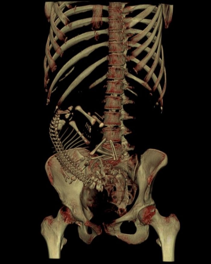
"This patient was involved in a motor vehicle accident, while 35 weeks pregnant.
Although exposure to the gravid uterus is to be avoided when ever possible, and only deliberately performed after careful weighing up of the pros and cons, McCollough et al conclude their recent Radiographics article with: “After comparing the doses from radiologic and nuclear medicine examinations with risk data from human in utero exposures, we have concluded that the absolute risks of fetal effects, including childhood cancer induction, are small at conceptus doses of 100 mGy and negligible at doses of less than 50mGy.” (1) Doses from diagnostic procedures are all below these thresholds - even a CT abdomen / pelvis only delivers approximately 30 mGy."
--Mark Hill 04:20, 24 August 2010 (UTC) - Timing may be Clinical (from LMP) rather than Embryological (from fertilization).
Image Source: http://www.radpod.org/2007/08/05/foetal-dosimetry/
Credit: Dr Frank Gaillard
This work is under a Creative Commons License
File history
Click on a date/time to view the file as it appeared at that time.
| Date/Time | Thumbnail | Dimensions | User | Comment | |
|---|---|---|---|---|---|
| current | 14:19, 24 August 2010 | | 700 × 874 (62 KB) | S8600021 (talk | contribs) | Text below from [http://www.radpod.org/ Radiology Picture of Day] "This patient was involved in a motor vehicle accident, while 35 weeks pregnant. Although exposure to the gravid uterus is to be avoided when ever possible, and only deliberately perform |
You cannot overwrite this file.
File usage
There are no pages that use this file.