File:Gilbert1957 fig02.jpg

Original file (1,280 × 824 pixels, file size: 130 KB, MIME type: image/jpeg)
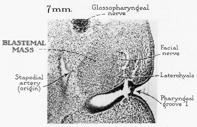
Fig. 2. Diagrams, based on reconstructions of embryos of horizons xv, xvi, and xvii
Illustrating the relations of the derivatives of the maxillomandibular mesoderm (figs. A, D) and the premantlibular condensation (figs. B, E) to the brain, eye vesicle, Gasserian ganglion, and mandibular arch. figure C represents a transection through the brain of an embryo of horizon xv at the level indicated in figure A. The derivatives of the maxillomandibular mesoderm and the premandibular condensation have been superimposed on the left side; the developing eye is illustrated on the right side of the transection.
Reference
Gilbert PW. The origin and development of the human extrinsic ocular muscles. (1957) Carnegie Instn. Wash. Publ. 611, Contrib. Embryol., Carnegie Inst. Wash. 36: 59-78.
Cite this page: Hill, M.A. (2024, June 26) Embryology Gilbert1957 fig02.jpg. Retrieved from https://embryology.med.unsw.edu.au/embryology/index.php/File:Gilbert1957_fig02.jpg
- © Dr Mark Hill 2024, UNSW Embryology ISBN: 978 0 7334 2609 4 - UNSW CRICOS Provider Code No. 00098G
File history
Yi efo/eka'e gwa ebo wo le nyangagi wuncin ye kamina wunga tinya nan
| Gwalagizhi | Nyangagi | Dimensions | User | Comment | |
|---|---|---|---|---|---|
| current | 13:52, 1 January 2018 |  | 1,280 × 824 (130 KB) | Z8600021 (talk | contribs) | |
| 13:52, 1 January 2018 |  | 1,900 × 1,876 (360 KB) | Z8600021 (talk | contribs) | Fig. 2. Diagrams, based on reconstructions of embryos of horizons xv, xvi, and xvii, illustrating the relations of the derivatives of the maxillomandibular mesoderm (figs. A, D) and the premantlibular condensation (figs. B, E) to the brain, eye vesicle... |
You cannot overwrite this file.
File usage
The following file is a duplicate of this file (more details):
The following page uses this file: