File:Cardiac muscle histology.jpg
Cardiac_muscle_histology.jpg (300 × 400 pixels, file size: 42 KB, MIME type: image/jpeg)
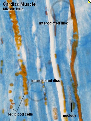
Cardiac Muscle, primate - Alizarin blue Use a low magnification to find a part of the tissue in which the cardiac muscle cells are cut longitudinally. At high magnification you should see striations and the large nuclei of the cardiac muscle cells. If you follow the course of individual cardiac muscle cells you will note fine, dark blue lines which seem to cross the fibres. These are the intercalated discs which connect the individual muscle cells and permit the conduction of electrical impulses between the cells
Cam40ab.jpg http://www.lab.anhb.uwa.edu.au/mb140/CorePages/Muscle/Images/cam40ab.jpg
Source: UWA Blue Histology
File history
Yi efo/eka'e gwa ebo wo le nyangagi wuncin ye kamina wunga tinya nan
| Gwalagizhi | Nyangagi | Dimensions | User | Comment | |
|---|---|---|---|---|---|
| current | 00:17, 17 August 2009 | | 300 × 400 (42 KB) | S8600021 (talk | contribs) | Cardiac Muscle, primate - Alizarin blue Use a low magnification to find a part of the tissue in which the cardiac muscle cells are cut longitudinally. At high magnification you should see striations and the large nuclei of the cardiac muscle cells. If you |
You cannot overwrite this file.
File usage
The following 11 pages use this file:
- ANAT2241 Cardiovascular System
- ANAT2241 Muscle Tissue
- Cardiac Muscle Histology
- Cardiovascular - Venous Development
- Cardiovascular System - Heart Histology
- Cardiovascular System Development
- Foundations - Histology Cells and Tissues
- HM Practical - Cardiac Histology
- Histology
- Histology Stains
- Muscle Development


