File:Mouse 3D Heart internal E8.5-14.5.jpeg
Original file (1,280 × 599 pixels, file size: 106 KB, MIME type: image/jpeg)
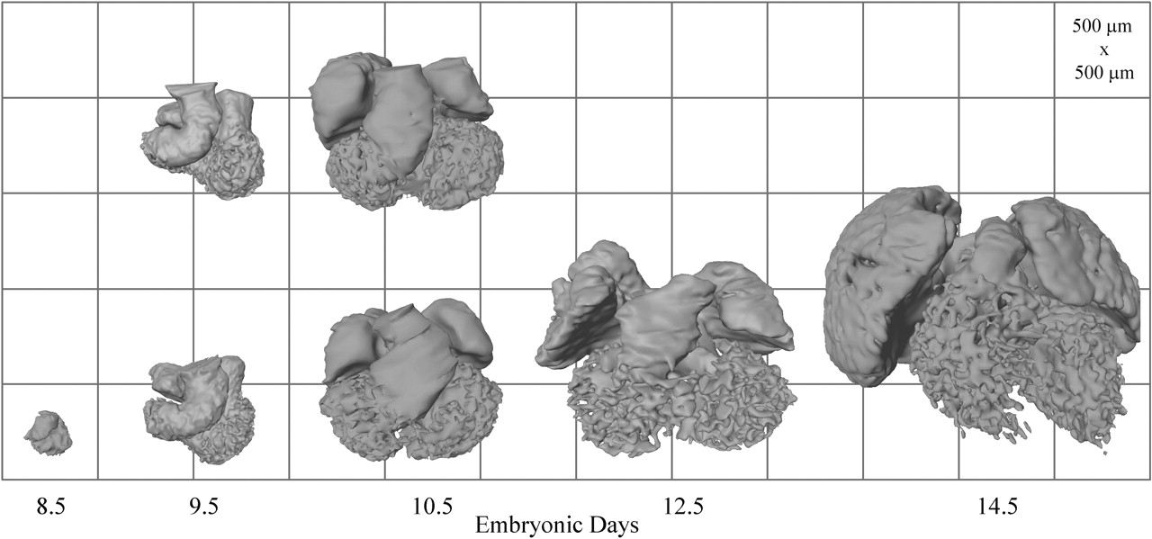
Reconstructions of the lumen of the hearts shown in external view. Duplicate reconstructions are shown for ED 9.5 and 10.5. This series clearly shows the formation of atrial (smooth walled) and ventricular (trabeculated) lumen from the outer curvature of the heart tube.
h71230363005.jpeg
Image (used with permission) from the paper Soufan AT, Ruijter JM, van den Hoff MJ, de Boer PA, Hagoort J, Moorman AF. Three-dimensional reconstruction of gene expression patterns during cardiac development. Physiol Genomics. 2003 May 13;13(3):187-95. (Physiol Genomics paper)
File history
Yi efo/eka'e gwa ebo wo le nyangagi wuncin ye kamina wunga tinya nan
| Gwalagizhi | Nyangagi | Dimensions | User | Comment | |
|---|---|---|---|---|---|
| current | 20:51, 16 August 2009 | | 1,280 × 599 (106 KB) | S8600021 (talk | contribs) | Reconstructions of the lumen of the hearts shown in external view. Duplicate reconstructions are shown for ED 9.5 and 10.5. This series clearly shows the formation of atrial (smooth walled) and ventricular (trabeculated) lumen from the outer curvature of |
You cannot overwrite this file.
File usage
The following 3 pages use this file:


