File:Hertig1945d plate01.jpg
Original file (1,280 × 2,002 pixels, file size: 427 KB, MIME type: image/jpeg)
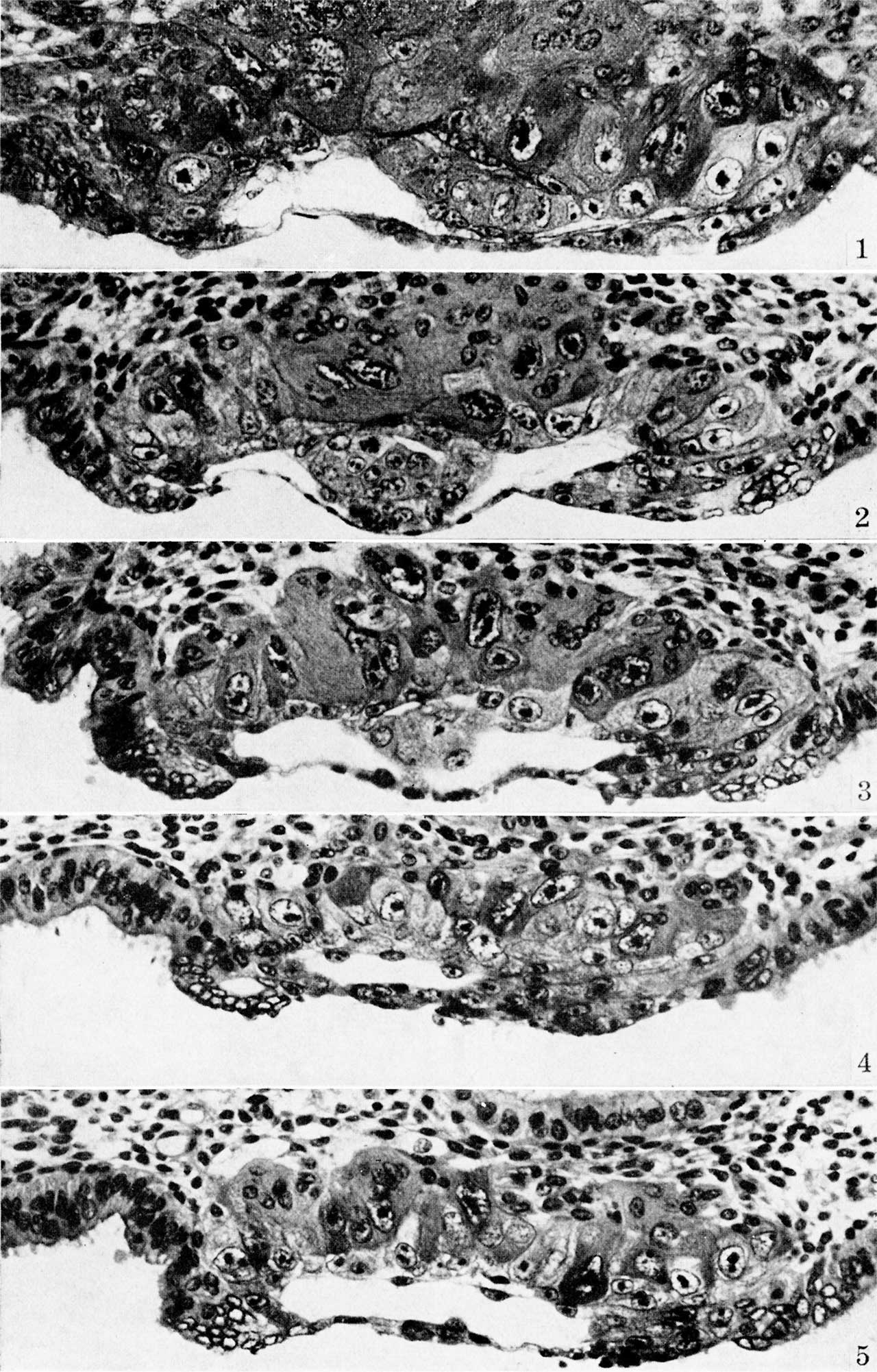
Plate 1. Ova of the eighth day
Fig. 1. The middle cross section of an ovum not over 7.5 days of age. Above the somewhat eccentrically placed germ-disk is a barely discernible cleft—the amniotic cavity. Above the latter are a few dark, flattened amniogenic cells de-lamiznating in situ from the adjacent trophoblast. Toward the left of the chorionic cavity, a few primitive mesoblastic cells are seen which likewise arise from trophoblast by in situ delamination. Carnegie No. 8225, section 20-5-5, X 300 (minus).
Fig. 2. The middle cross section of a 7.5-day ovum. The cleft-like amniotic cavity lies above the germ-disk and beneath amniogenic cells arising from the adjacent trophoblast. A single, flattened meso-blastic cell is seen to the right of the germ-disk and lying upon the inner aspect of the tropho-blast. Carnegie No. 8020, section 6-5-9, X 300 (minus).
Fig. 3. The same ovum as fig. 2 but 7 sections removed. The amniotic cavity is less evident because of its smaller size although the amniogenic cells overlying it are more prominent. Carnegie No. 8020, section 6-5-2, X 300.
Fig. 4. The same ovum as shown in figs. 2 and 3 but the section passes to one side of the germ-disk. Note at the embryonic pole the three prominent mesoblastic cells which are delaminating in situ from the adjacent troplgoblast. A somewhat more advanced stage of this process is seen in the two mesoblastic cells shown in fig. 5. Carnegie 8020, section 6-3-6, X 300 (plus).
Fig. 5. A section of a. 7.5-day ovum (same specimen as figs. 2-4) showing a more advanced stage of mesoblastic formation. Note the two, nearly detached mesoblastic cells delaminating from, but still lightly attached to the trophoblast at the embryonic pole of the ovum. Carnegie 8020, section 6-4-3, X 300 (plus).
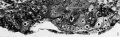
Fig 1 Carnegie No. 8225
Fig 2 Carnegie No. 8020
Fig 3 Carnegie No. 8020
Fig 4 Carnegie No. 8020
Fig 5 Carnegie No. 8020
Reference
Hertig AT. On the development of the amnion and exocoelomic membrane in the previllous human ovum. (1945) Yale J Biol Med. 18:107-15. PubMed 21007544
Cite this page: Hill, M.A. (2024, May 23) Embryology Hertig1945d plate01.jpg. Retrieved from https://embryology.med.unsw.edu.au/embryology/index.php/File:Hertig1945d_plate01.jpg
- © Dr Mark Hill 2024, UNSW Embryology ISBN: 978 0 7334 2609 4 - UNSW CRICOS Provider Code No. 00098G
File history
Click on a date/time to view the file as it appeared at that time.
| Date/Time | Thumbnail | Dimensions | User | Comment | |
|---|---|---|---|---|---|
| current | 13:34, 24 October 2017 | | 1,280 × 2,002 (427 KB) | Z8600021 (talk | contribs) | |
| 13:34, 24 October 2017 | 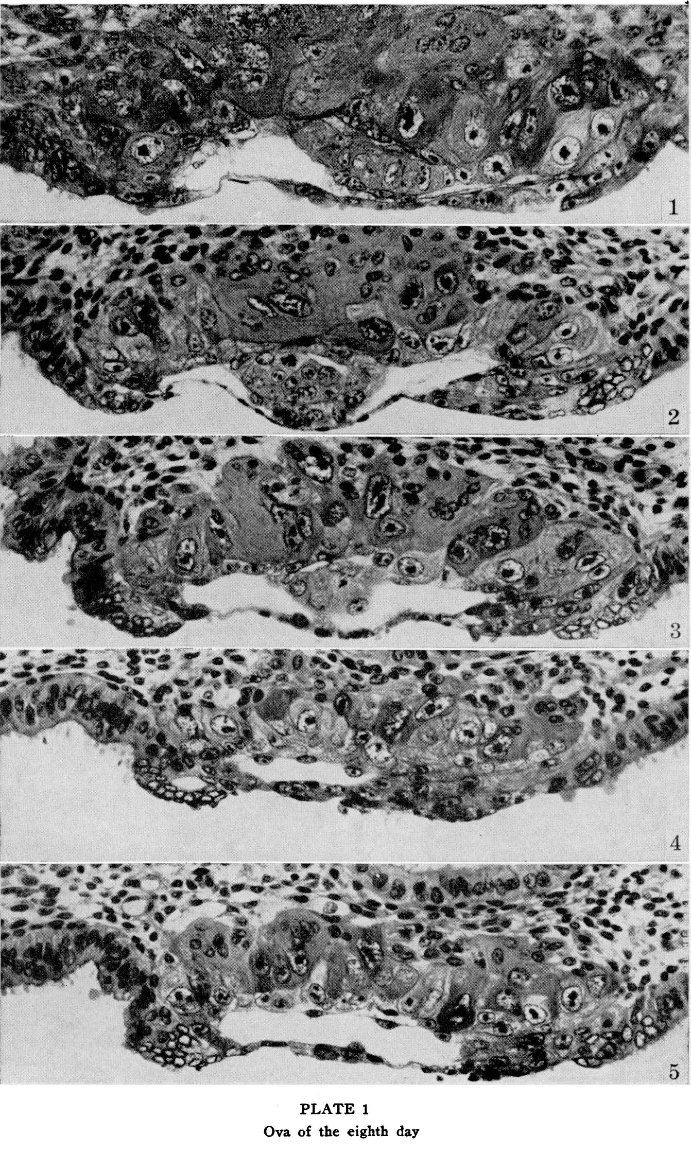 | 1,342 × 2,240 (726 KB) | Z8600021 (talk | contribs) | ==Plate 1. Ova of the eighth day== Fig. 1. The middle cross section of an ovum not over 7% days of age. Above the somewhat eccentrically placed germ-disk is a barely discernible cleft—the amniotic cavity. Above the latter are a few dark, flattened... |
You cannot overwrite this file.
File usage
The following page uses this file: