File:Boyden1931 fig05.jpg

Original file (1,000 × 646 pixels, file size: 97 KB, MIME type: image/jpeg)
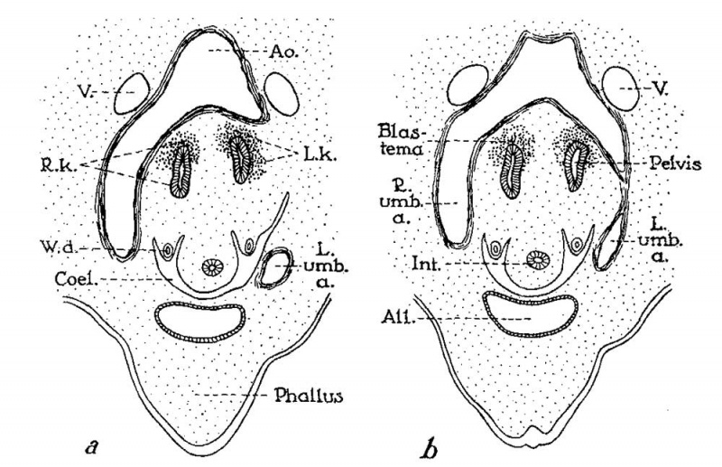
Fig. 5. Cross-sections through 11.5 mm human embryo
(age, thirty-two to thirty-three days), illustrating normal position of kidneys in crotch of umbilical arteries. a,b (two sections apart) from text figures 29 and 30 of embryo “H. s. Bul.” (after Keibel, ’96). Note that pelvis of each kidney is directed anteriorly, an unrotated position that is retained in most cases of horseshoe kidney.
All, allantois; Ao., aorta; Coel., body cavity; Int, intestine; L. and R. umb.a., left and right umbilical arteries; W.d., Wolffian duct.
Reference
Boyden EA. Description of a horseshoe kidney associated with left inferior vena cava and disc-shaped suprarenal glands, together with a note on the occurrence of horseshoe kidneys in human embryos. (1931) Anat. Rec. 51(2): 187-211.
Cite this page: Hill, M.A. (2024, June 10) Embryology Boyden1931 fig05.jpg. Retrieved from https://embryology.med.unsw.edu.au/embryology/index.php/File:Boyden1931_fig05.jpg
- © Dr Mark Hill 2024, UNSW Embryology ISBN: 978 0 7334 2609 4 - UNSW CRICOS Provider Code No. 00098G
File history
Click on a date/time to view the file as it appeared at that time.
| Date/Time | Thumbnail | Dimensions | User | Comment | |
|---|---|---|---|---|---|
| current | 13:52, 8 September 2017 | | 1,000 × 646 (97 KB) | Z8600021 (talk | contribs) | |
| 13:51, 8 September 2017 |  | 1,325 × 1,032 (216 KB) | Z8600021 (talk | contribs) |
You cannot overwrite this file.
File usage
The following page uses this file: