File:Hertig1946b fig25.jpg
Original file (800 × 1,184 pixels, file size: 266 KB, MIME type: image/jpeg)
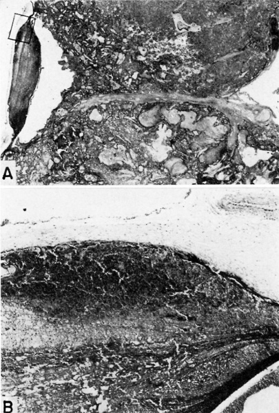
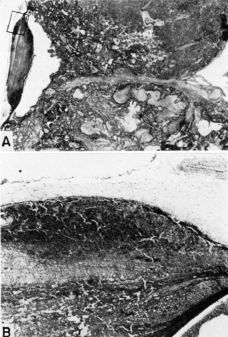
Fig. 25. A human placenta at term to show diffuse and discrete thrombosis of intervillous space. B. L.—i. H., S-32-217.
A. Laminated fibrin thrombus deposited on under surface of chorionic membrane at left of picture. Note septum running horizontally between two adjacent cotyledons, the lower of which is infarcted due to diffuse and discrete intervillous thrombosis while the top one is normal. X5.
B. A more highly magnified view of rectangle seen in A. The fibrous tissue of the chorionic membrane is at the top, beneath which is the cytotrophoblast whose “fibrinoid” degeneration has caused the discrete mass of fibrin to be deposited from the maternal blood. X60.
File history
Yi efo/eka'e gwa ebo wo le nyangagi wuncin ye kamina wunga tinya nan
| Gwalagizhi | Nyangagi | Dimensions | User | Comment | |
|---|---|---|---|---|---|
| current | 13:04, 8 August 2017 | | 800 × 1,184 (266 KB) | Z8600021 (talk | contribs) | Fig. 25. A human placenta at term to show diffuse and discrete thrombosis of intervillous space. B. L.—i. H., S-32-217. A. Laminated fibrin thrombus deposited on under surface of chorionic membrane at left of picture. Note septum running horizontal... |
You cannot overwrite this file.
File usage
The following page uses this file: