File:Hertig1946b fig05b.jpg
Original file (800 × 627 pixels, file size: 116 KB, MIME type: image/jpeg)
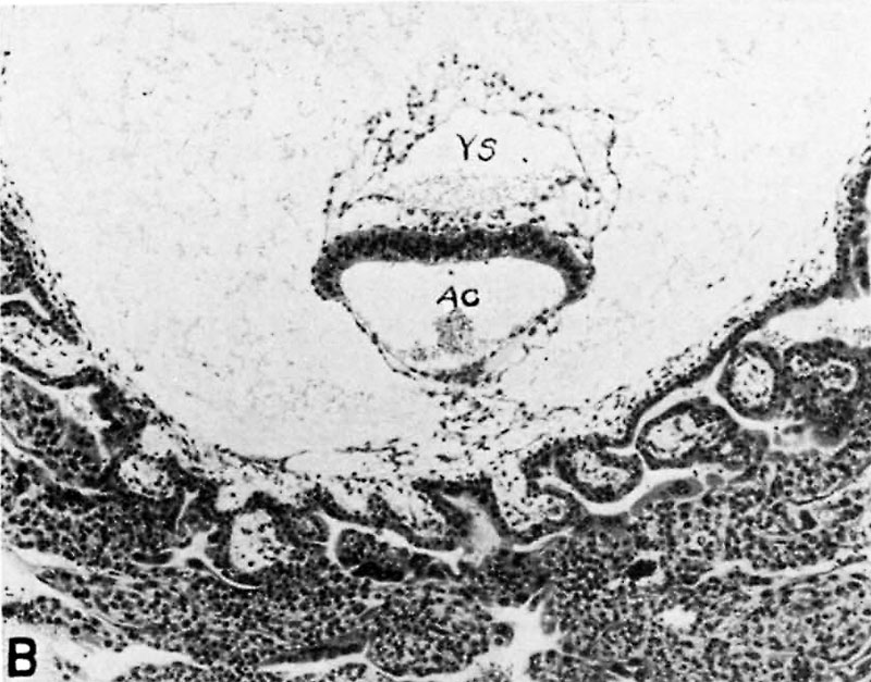
Fig. 5 B. Section through embryo of 16.5 day human ovum.
Section through embryo of 16.5 day human ovum. Note double-layered yolk-sac (YS) above and amniotic cavity (AC) below the embryonic disk. The amnion is lightly attached by mesoblastic tissue to the chorionic membrane whose simple villi are continuous with their trophoblastic cell columns. The latter are continuous with the surrounding endometrium although this feature is not seen in this picture. Carnegie 7802, section 44-3-5, X100.
References
Hertig AT. lnvolution of tissues in fetal life: a review. (1946) Anat. Rec. 94: 96-116.
Cite this page: Hill, M.A. (2024, May 23) Embryology Hertig1946b fig05b.jpg. Retrieved from https://embryology.med.unsw.edu.au/embryology/index.php/File:Hertig1946b_fig05b.jpg
- © Dr Mark Hill 2024, UNSW Embryology ISBN: 978 0 7334 2609 4 - UNSW CRICOS Provider Code No. 00098G
File history
Click on a date/time to view the file as it appeared at that time.
| Date/Time | Thumbnail | Dimensions | User | Comment | |
|---|---|---|---|---|---|
| current | 16:54, 7 August 2017 | | 800 × 627 (116 KB) | Z8600021 (talk | contribs) |
You cannot overwrite this file.
File usage
The following 2 pages use this file: