File:GladstoneHamilton1941 text-fig09.jpg

Original file (1,280 × 638 pixels, file size: 89 KB, MIME type: image/jpeg)
Text-fig. 9. A section just behind the anterior intestinal portal
The lateral infoldings of the yolk sac wall, and the marginal clefts in the mesenchyme of the embryonic plate in the region of the pericardium, are visible. The medullary plate which in Text-figs. 7 and 8 consists of a single layer of flattened eotodermal cells is now considerably thicker and two or three layers of nuclei are visible. Section no. 12. . x 100.
Reference
Gladstone RJ. and Hamilton WJ. A presomite human embryo (Shaw) with primitive streak and chorda canal with special reference to the development of the vascular system. (1941) Amer. J Anat. 76(1): 9-44.
Cite this page: Hill, M.A. (2026, October 4) Embryology GladstoneHamilton1941 text-fig09.jpg. Retrieved from https://embryology.med.unsw.edu.au/embryology/index.php/File:GladstoneHamilton1941_text-fig09.jpg
- © Dr Mark Hill 2026, UNSW Embryology ISBN: 978 0 7334 2609 4 - UNSW CRICOS Provider Code No. 00098G
File history
Yi efo/eka'e gwa ebo wo le nyangagi wuncin ye kamina wunga tinya nan
| Gwalagizhi | Nyangagi | Dimensions | User | Comment | |
|---|---|---|---|---|---|
| current | 18:12, 26 February 2017 | | 1,280 × 638 (89 KB) | Z8600021 (talk | contribs) | |
| 18:12, 26 February 2017 | 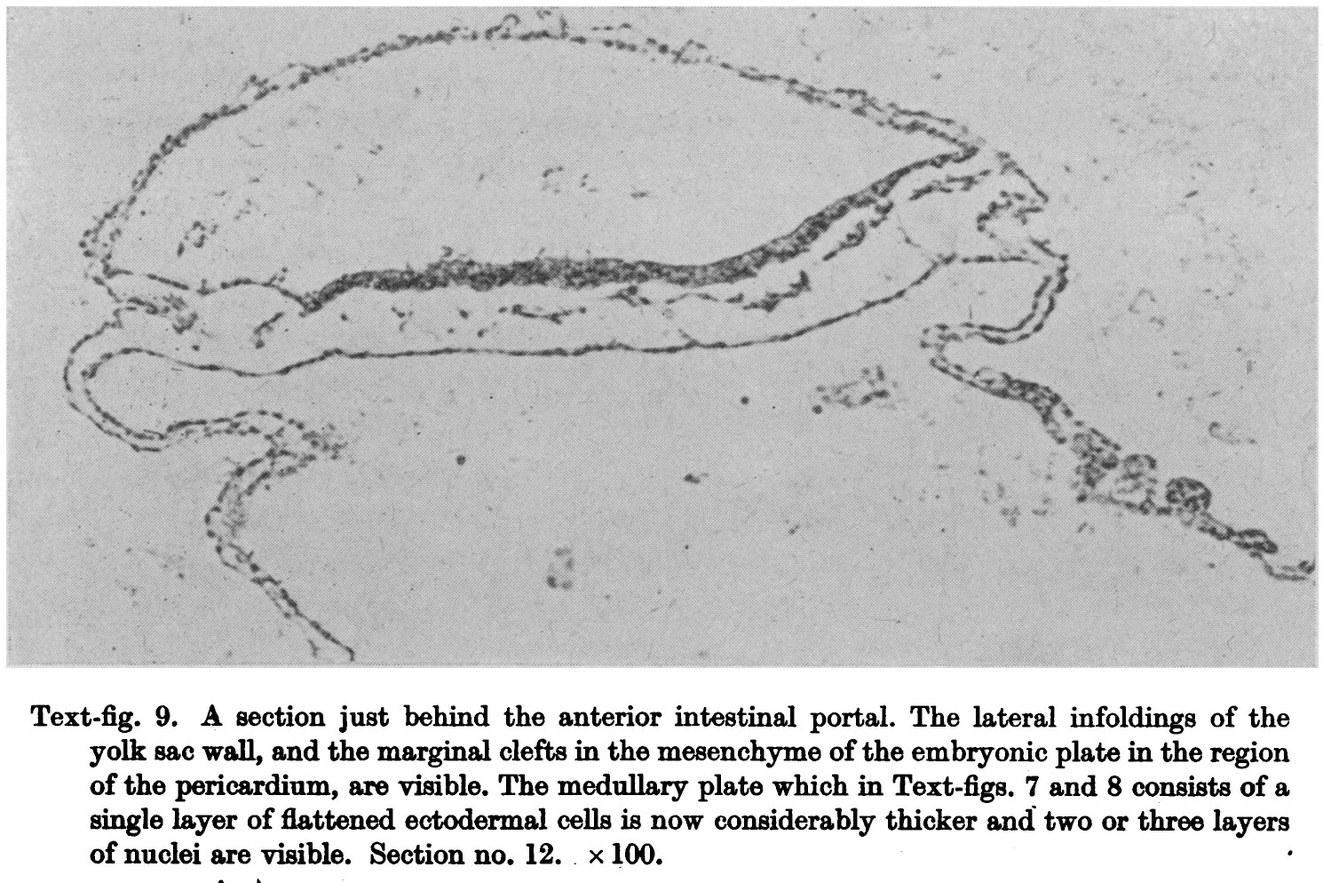 | 1,490 × 994 (368 KB) | Z8600021 (talk | contribs) |
You cannot overwrite this file.


