File:GladstoneHamilton1941 text-fig08.jpg
Original file (1,000 × 693 pixels, file size: 120 KB, MIME type: image/jpeg)
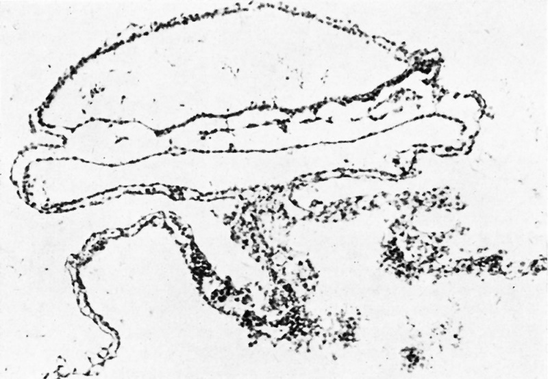
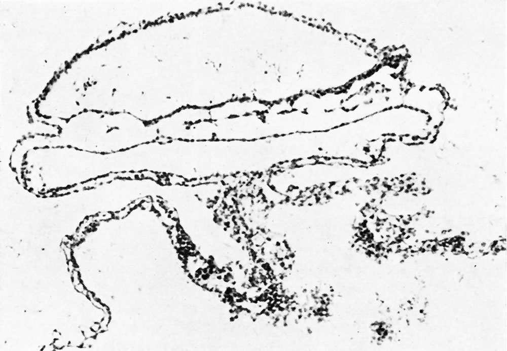
Text-fig. 8. The section passes through the anterior boundary of the anterior intestinal--portal
Shows the junction of the lateral folds of the yolk sac wall. Blood islands and empty endothelial-‘lined spaces are present in the wall of the yolk sac, andin the floor and roof of the foregut. Section no. 10. x 100.
Reference
Gladstone RJ. and Hamilton WJ. A presomite human embryo (Shaw) with primitive streak and chorda canal with special reference to the development of the vascular system. (1941) Amer. J Anat. 76(1): 9-44.
Cite this page: Hill, M.A. (2026, October 3) Embryology GladstoneHamilton1941 text-fig08.jpg. Retrieved from https://embryology.med.unsw.edu.au/embryology/index.php/File:GladstoneHamilton1941_text-fig08.jpg
- © Dr Mark Hill 2026, UNSW Embryology ISBN: 978 0 7334 2609 4 - UNSW CRICOS Provider Code No. 00098G
File history
Yi efo/eka'e gwa ebo wo le nyangagi wuncin ye kamina wunga tinya nan
| Gwalagizhi | Nyangagi | Dimensions | User | Comment | |
|---|---|---|---|---|---|
| current | 18:09, 26 February 2017 | | 1,000 × 693 (120 KB) | Z8600021 (talk | contribs) | |
| 18:09, 26 February 2017 |  | 1,447 × 1,131 (373 KB) | Z8600021 (talk | contribs) |
You cannot overwrite this file.