File:Anderson2016-fig01.jpg
From Embryology

Size of this preview: 600 × 600 pixels.
Original file (800 × 800 pixels, file size: 93 KB, MIME type: image/jpeg)
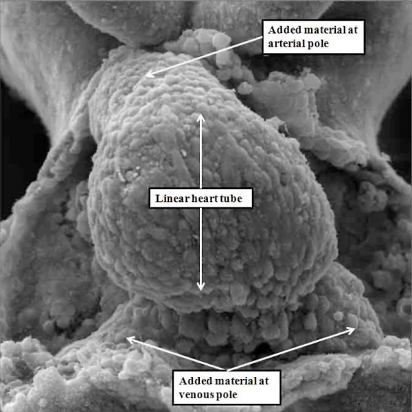
Fig. 1. Mouse E8 linear heart tube (SEM)
The image is a scanning electron micrograph showing the linear heart tube of the mouse within the pericardial cavity.
Reference
Anderson RH. Teratogenecity in the setting of cardiac development and maldevelopment. (2016)
Cite this page: Hill, M.A. (2024, June 26) Embryology Anderson2016-fig01.jpg. Retrieved from https://embryology.med.unsw.edu.au/embryology/index.php/File:Anderson2016-fig01.jpg
- © Dr Mark Hill 2024, UNSW Embryology ISBN: 978 0 7334 2609 4 - UNSW CRICOS Provider Code No. 00098G
File history
Yi efo/eka'e gwa ebo wo le nyangagi wuncin ye kamina wunga tinya nan
| Gwalagizhi | Nyangagi | Dimensions | User | Comment | |
|---|---|---|---|---|---|
| current | 13:47, 16 February 2017 | | 800 × 800 (93 KB) | Z8600021 (talk | contribs) | ===Reference=== {{Ref-Anderson2016}} {{Footer}} |
You cannot overwrite this file.
File usage
The following 3 pages use this file: