File:McMurrich1914 fig279.jpg
From Embryology
Size of this preview: 507 × 600 pixels. Other resolution: 845 × 1,000 pixels.
Original file (845 × 1,000 pixels, file size: 176 KB, MIME type: image/jpeg)
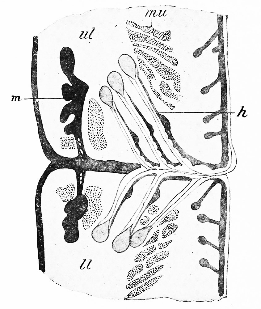
Fig. 279. Section through the Margins of the Fused Eyelids in an Embryo of Six Months
ih, Eyelash; //, lower lid; m, tarsal gland; mu, muscle bundle; ul, upper lid. Seidl.)
-(Schweigger
| Historic Disclaimer - information about historic embryology pages |
|---|
 |
Reference
McMurrich JP. The Development Of The Human Body. (1914) P. Blakiston's Son & Co., Philadelphia, Pennsylvania.
Cite this page: Hill, M.A. (2024, June 17) Embryology McMurrich1914 fig279.jpg. Retrieved from https://embryology.med.unsw.edu.au/embryology/index.php/File:McMurrich1914_fig279.jpg
- © Dr Mark Hill 2024, UNSW Embryology ISBN: 978 0 7334 2609 4 - UNSW CRICOS Provider Code No. 00098G
File history
Yi efo/eka'e gwa ebo wo le nyangagi wuncin ye kamina wunga tinya nan
| Gwalagizhi | Nyangagi | Dimensions | User | Comment | |
|---|---|---|---|---|---|
| current | 22:27, 31 January 2017 | | 845 × 1,000 (176 KB) | Z8600021 (talk | contribs) | |
| 22:25, 31 January 2017 | 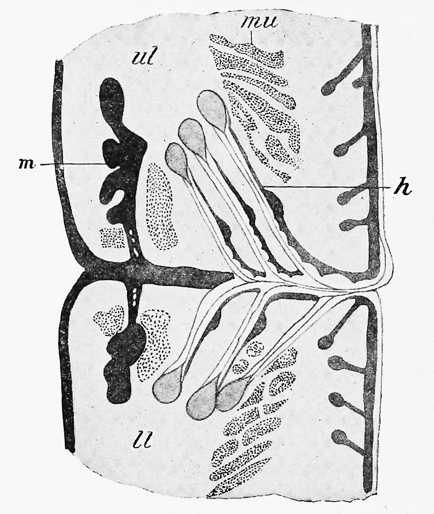 | 845 × 1,000 (191 KB) | Z8600021 (talk | contribs) | ||
| 22:23, 31 January 2017 | 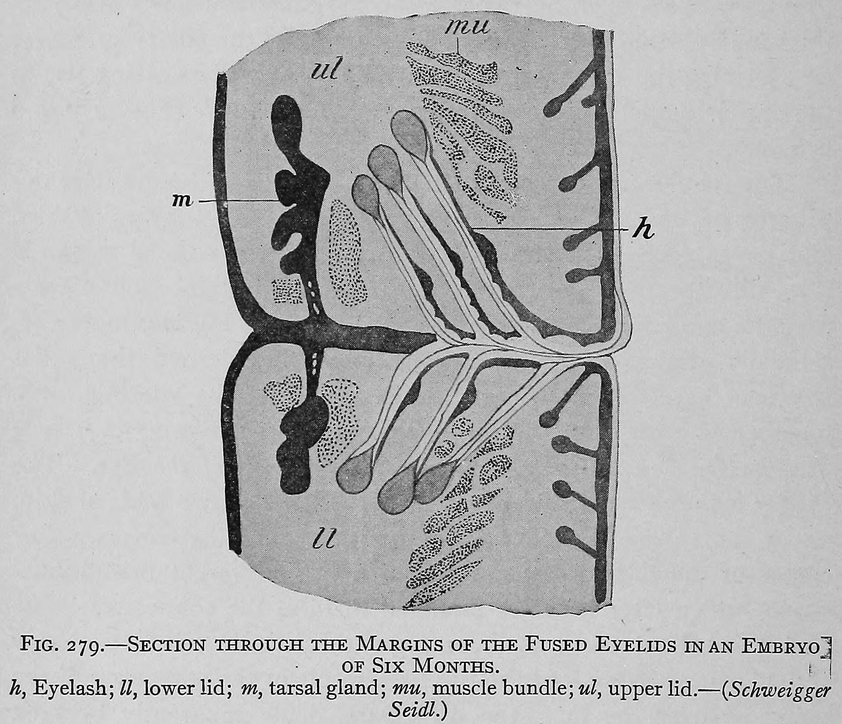 | 1,713 × 1,474 (482 KB) | Z8600021 (talk | contribs) |
You cannot overwrite this file.
File usage
The following page uses this file: