Paper - The development of the hypophysis cerebri of the rabbit
| Embryology - 16 Jun 2024 |
|---|
| Google Translate - select your language from the list shown below (this will open a new external page) |
|
العربية | català | 中文 | 中國傳統的 | français | Deutsche | עִברִית | हिंदी | bahasa Indonesia | italiano | 日本語 | 한국어 | မြန်မာ | Pilipino | Polskie | português | ਪੰਜਾਬੀ ਦੇ | Română | русский | Español | Swahili | Svensk | ไทย | Türkçe | اردو | ייִדיש | Tiếng Việt These external translations are automated and may not be accurate. (More? About Translations) |
Atwell WJ. The development of the hypophysis cerebri of the rabbit (Lepus Cuniculus L.). (1918) Amer. J Anat. 24(2): 271-337
| Online Editor |
|---|
| Wayne J. Attwell (1889 - 1941) student of GC. Huber.
See also Atwell WJ. The development of the hypophysis cerebri in man, with special reference to the pars tuberalis. (1926) Amer. J Anat. 37: 139-193. Links: Pituitary Development | Rabbit Development |
| Historic Disclaimer - information about historic embryology pages |
|---|
| Pages where the terms "Historic" (textbooks, papers, people, recommendations) appear on this site, and sections within pages where this disclaimer appears, indicate that the content and scientific understanding are specific to the time of publication. This means that while some scientific descriptions are still accurate, the terminology and interpretation of the developmental mechanisms reflect the understanding at the time of original publication and those of the preceding periods, these terms, interpretations and recommendations may not reflect our current scientific understanding. (More? Embryology History | Historic Embryology Papers) |
The Development of the Hypophysis Cerebri of the Rabbit (Lepus Cuniculus L.)
1. Introduction
2. Review of literature
- A. Special questions concerned with the development of the hypophysis
- 1. Entodermal origin
- 2. The relation of the notochord to the hypophysis
- 3. The lobes of the hypophysis
- a) General
- b) The anterior process
- c) The lateral lobes
- d) The relation of the lateral lobes to the 'pars tuberalis'
- 4. The structure of the intermediate part
- 5. The development of the neural lobe
- B. The development of the hypophysis of the rabbit
3. Material and methods
4. The development of the hypophysis of the rabbit by days
5. Discussion of observations
- A. The relation of the entoderm and the notochord to the hypophysis
- B. Some general features of the development of the hypophysis
- 1. The hypophysial stalk
- 2. The residual lumen
- D. The development of the neural lobe
- E. The neuro-epithelial contacts and their significance
- F. Terminology and phylogeny of the lobes of the hypophysis
6. Summary and conclusions
7. Literature cited
Introduction
Notwithstanding the numerous studies to which the hypophysis has been subjected, many of its deeper problems remain unsolved. Few investigators have confined themselves to the development of the gland in a single species. This is due, no doubt, to the alluring possibilities in broad comparative studies. As a consequence, many sweeping and unwarranted conclusions have been drawn from insufiicient observations, or from observations on a few specimens in widely different vertebrate classes.
The recent recognition of a distinctive third epithelial portion of the gland lying under the membranes of the brain in the region of the tuber cinereum—the ‘pars tuberalis’ of Tilney (’l3)—and the as yet imperfect appreciation of its interesting development, make careful ontogcnctic studies highly desirable as the basis for phylogenetic comparisons.
The Work which forms the basis of the present paper was undertaken in an attempt to trace the development of the hypophysis with reasonable completeness in a single mammal. To this end the rabbit (Lepus cuniculus L.) was chosen, since this animal breeds Well in confined quarters, has a short gestation period, and brings forth its young in large litters. These factors combine to make possible the collection of the carefully timed embryological material so essential to chronological studies in development.
This study will treat particularly of the morphogenesis of the hypophysis from the time of its appearance until birth, giving especial attention to the ontogeny of the ‘pars tuberalis’ and to the development of the neural lobe; it will deal also with the differentiated histological structure of the three parts of the epithelial hypophysis at the time of birth, and finally will attempt to relate certain of the author’s observations to those of other investigators.
I desire to express my sincere thanks to Professor Huber for his continued interest in this Work, for his very material assistance in overcoming technical difficulties, and for the unstinting Way in which he has placed the excellent facilities of the anatomical laboratories at my disposal. My wife has given valuable aid in much of the tedious Work connected with the construction of the wax models.
Review of Literature
A. Special questions concerned with the development of the hypophysis
I . Entodermal origin. An endless chain of discussion has been evoked by the question as to whether the epithelial hypophysis is derived from the entoderm or from the ectoderm, or whether it is compounded of elements derived from both of these germ layers. Since the time of Goette C74), Balfour C74), and Mihalkovics C75), only a few authors have maintained that this portion of the gland is developed from entoderm alone. There have been many, however, to state that the primary ectodermal anlage is later augmented by a larger or smaller contribution from the entoderm.
Hoffman C86) and Ostroumoff C88) believe the hypophysis in certain reptiles to be entodermal in origin. Kupffer C94) describes a growth from the cephalic end of the foregut to meet the dorsal wall of Rathke’s pocket. This he interprets as an attempted recurrence of a primitive pre—oral mouth or ‘paleostoma.’ This suggestion of Kupffer’s has caused many writers to attach great phylogenetic importance to the hypophysis. Furthermore, according to Kupffer, the entoderm makes important contributions to the epithelial lobe of the hypophysis.
In a previous paper (Atwell, ’ 15) reference was made to the observations of Saint—Remy C95), Valenti C95 a, ’95 b, and ’97), Nusbaum C96), and of Bruni C14), according to all of whom the entoderrn of the pouch—like cephalic extremity of the foregut (‘Seessel’s pouch’) fuses with the ectodermal hypophysial anlage and possibly ‘contributes a few cells to it.
Orrù ('00) divides the glandular portion of the hypophysis of Gongylus ocellatus into two lobes, one of which is in relation to the infundibular process, While the other lies ventral to the first. Although he cannot trace a difference histologically in these two lobes, he believes it Very probable that the dorsal lobe, which lies close to the infundibular process is entodermal and that the ventral lobe is ectodermal.
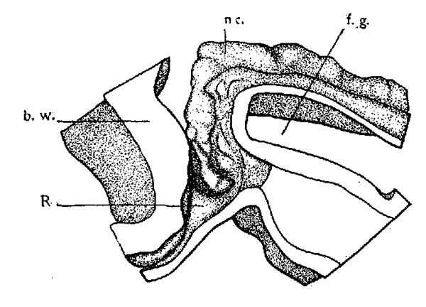
Fig. 1. Model of hypophysis region of a sixteen-somite rabbit (embryo A). x 100. The anterior end of the notochord and portions of the epithelium of the foregut, oral pit, and brain wall are shown. Viewed from the left side. nc., notochord; f.g., foregut; R,Rathka’s pouch, and b.w., brain wall.
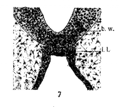
constriction from the oral epithelium. These prominent elevations I interpret as the homologues of the lateral lobes of lower
forms. Later stages of development of the rabbit will show how
these early lateral lobes give rise to the pars tuberalis of complete
development. A transverse section through the region of the
lateral lobes is shown in figure 7. The lobes are constricted from
the thickened epithelium just nasal to the early hypophyseal pouch.
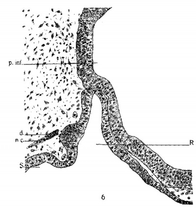
In embryo 25A (figs. 5 and 6) the notochord ends close to a large thickened evagination of the dorsal wall of the hypophysis from which it is separated by a very narrow space. The condition presented by this embryo corresponds very closely to that of the eleven-day rabbit embryo described by Koelliker (’79).
In embryo C, which is only slightly more advanced, the notochord divides into two parts near its cephalic termination. One branch ends at the apex of Seessel’s pouch, while the other is directed dorsally, forming almost a right angle with the first part.
| Historic Disclaimer - information about historic embryology pages |
|---|
| Pages where the terms "Historic" (textbooks, papers, people, recommendations) appear on this site, and sections within pages where this disclaimer appears, indicate that the content and scientific understanding are specific to the time of publication. This means that while some scientific descriptions are still accurate, the terminology and interpretation of the developmental mechanisms reflect the understanding at the time of original publication and those of the preceding periods, these terms, interpretations and recommendations may not reflect our current scientific understanding. (More? Embryology History | Historic Embryology Papers) |
Cite this page: Hill, M.A. (2024, June 16) Embryology Paper - The development of the hypophysis cerebri of the rabbit. Retrieved from https://embryology.med.unsw.edu.au/embryology/index.php/Paper_-_The_development_of_the_hypophysis_cerebri_of_the_rabbit
- © Dr Mark Hill 2024, UNSW Embryology ISBN: 978 0 7334 2609 4 - UNSW CRICOS Provider Code No. 00098G


