File:Atwell1926 plate07.jpg
Original file (1,000 × 1,033 pixels, file size: 104 KB, MIME type: image/jpeg)
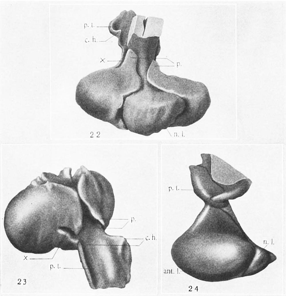
Plate 7 Wax-plate reconstruction of the hypophysis
22 Wax-plate reconstruction of the hypophysis from a 102-mm. human fetus (U.B.E.C., no. 17), viewed dorsally and from above. X 35.
23 The same model as shown in figure 22 with the neural lobe l‘(’Ill0\’('(l, vix-we<l from above and in front. X 35.
24 R4-vonstrtwtion of tho lxypophyeis from 9. human fetus of 28.5 cm. (l.T.B.l'}.(?., no. 45), viewed from the left side. X 15.
Abbreviations
rm.I.t., anterior lobe ' 1)., process surrounding neck of neural c.h. «caudally directed horn of para t.u- Iobo lneralis p.t., pars tuberalis n..l., neural lobe 3'-., incisure separating pars inte-rnu.-diu and pars tuberalis
Reference
Atwell WJ. The development of the hypophysis cerebri in man, with special reference to the pars tuberalis. (1926) Amer. J Anat. 37: 139-193.
File history
Yi efo/eka'e gwa ebo wo le nyangagi wuncin ye kamina wunga tinya nan
| Gwalagizhi | Nyangagi | Dimensions | User | Comment | |
|---|---|---|---|---|---|
| current | 14:57, 9 November 2016 | | 1,000 × 1,033 (104 KB) | Z8600021 (talk | contribs) | |
| 14:56, 9 November 2016 | 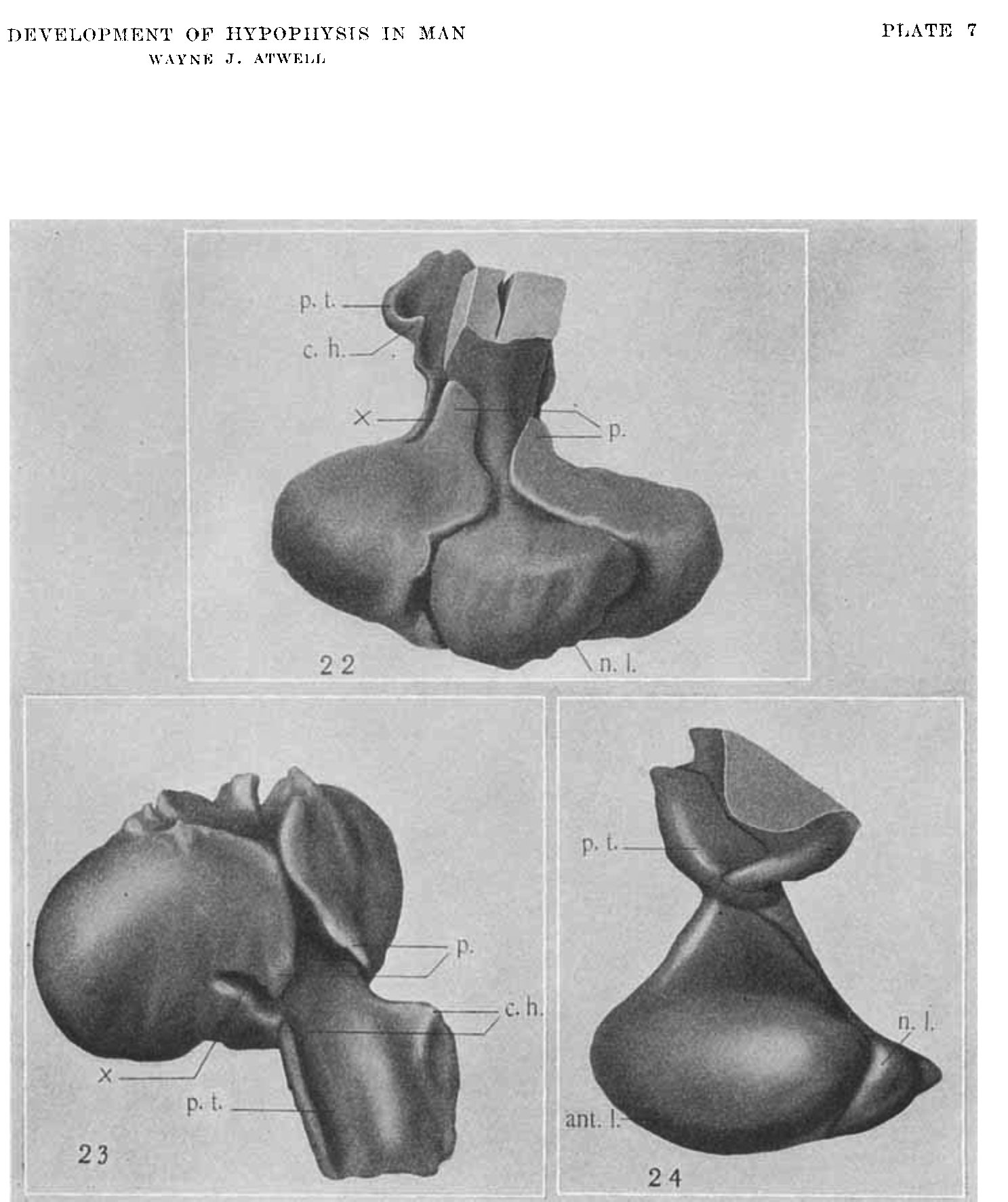 | 1,487 × 1,773 (240 KB) | Z8600021 (talk | contribs) | ===Reference=== {{Ref-Atwell1926}} |
You cannot overwrite this file.
File usage
The following page uses this file: