File:Nelsen1953 fig246.jpg

Original file (1,200 × 986 pixels, file size: 253 KB, MIME type: image/jpeg)
Fig. 246. Development of body form in human embryo
(C from Keibel and Mall: Manual of Human Embryology, Vol. I, 1910. Philadelphia and London, Lippincott. A, B, D, and E from Keibel and Elze: Normentafel zur Entwicklungsgeschkhte des Menschen. Jena, 1908. G. Fischer.)
(A) Early neural fold stage. Somites are beginning to form; notochordal canal is evident.
(B) About nine pairs of somites.
(C) His's embryo M.
(D) About 23 pairs of somites, 4-5 mm. long.
(E) About 35 pairs of trunk somites, 12 mm. long.
Reference
Nelsen OE. Comparative embryology of the vertebrates (1953) Mcgraw-Hill Book Company, New York.
Cite this page: Hill, M.A. (2024, June 15) Embryology Nelsen1953 fig246.jpg. Retrieved from https://embryology.med.unsw.edu.au/embryology/index.php/File:Nelsen1953_fig246.jpg
- © Dr Mark Hill 2024, UNSW Embryology ISBN: 978 0 7334 2609 4 - UNSW CRICOS Provider Code No. 00098G
File history
Click on a date/time to view the file as it appeared at that time.
| Date/Time | Thumbnail | Dimensions | User | Comment | |
|---|---|---|---|---|---|
| current | 14:37, 26 October 2016 | | 1,200 × 986 (253 KB) | Z8600021 (talk | contribs) | |
| 14:36, 26 October 2016 | 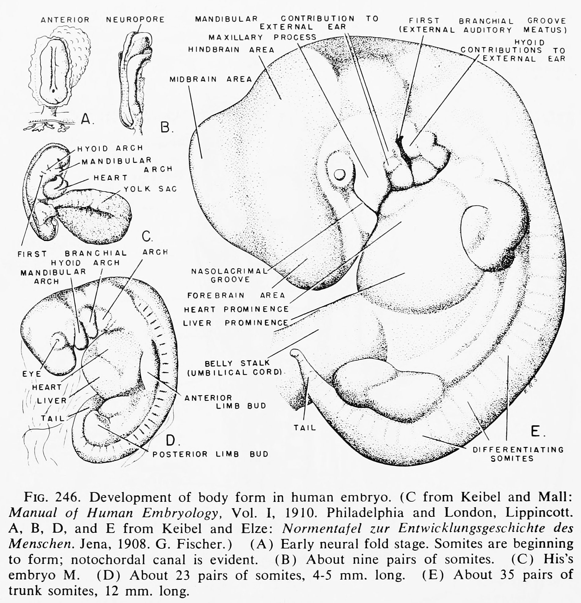 | 1,901 × 1,973 (645 KB) | Z8600021 (talk | contribs) |
You cannot overwrite this file.
File usage
The following 2 pages use this file: