File:Arey1924 fig197.jpg
Original file (1,200 × 1,615 pixels, file size: 227 KB, MIME type: image/jpeg)
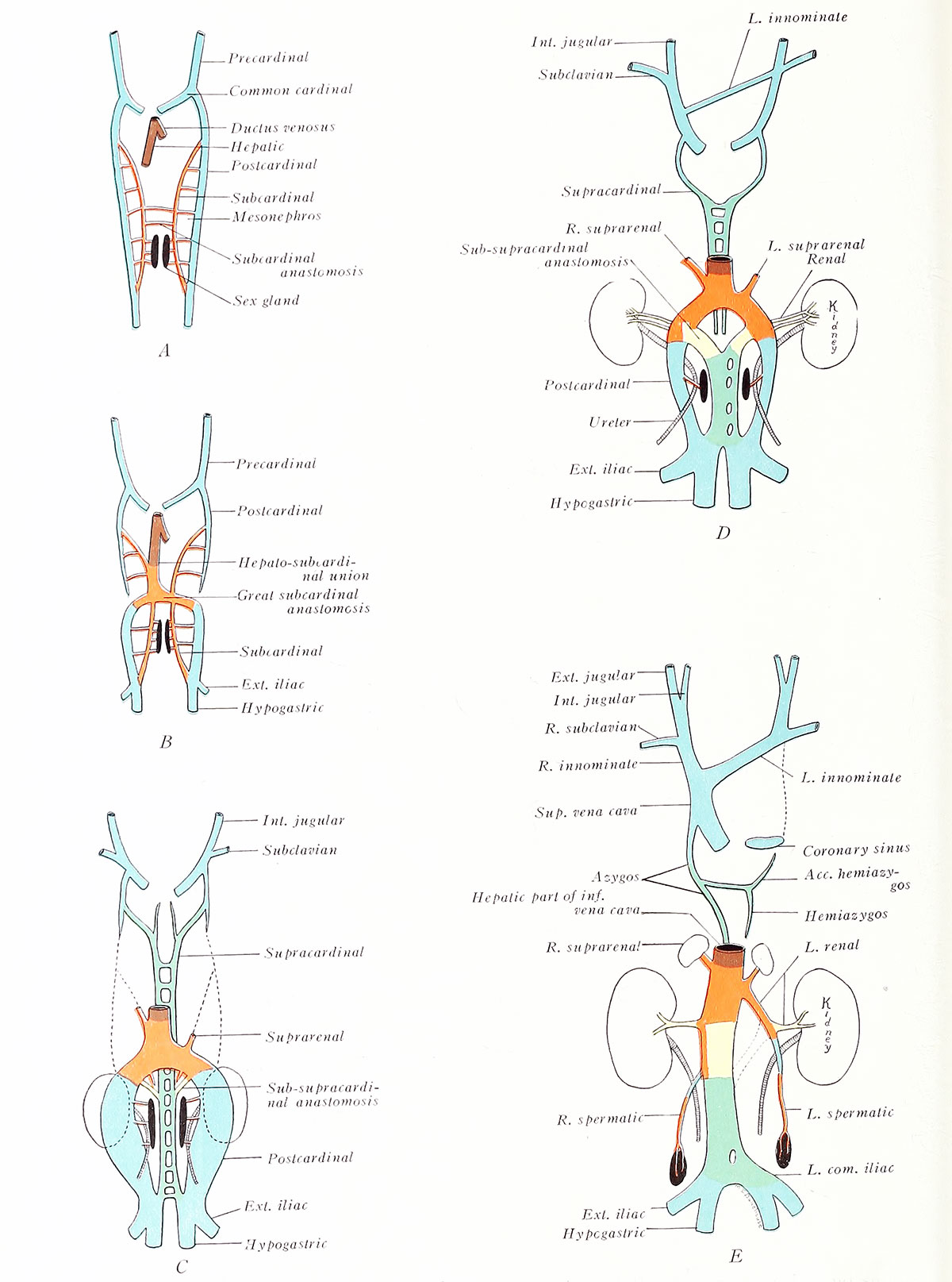
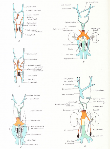
Fig. 197. Diagrams to illustrate the transformation of the pre-, post-, sub-, and supracardinal veins
(adapted after Huntington and McClure)
A, 6 mm; B, 10 mm; C,15 mm, D, 18 mm.
The true precardinals communicate during the eighth week by a transverse venous channel which carries the blood from the left side of the head into the right vein (Fig. 197 D). As a result, the left precardinal soon loses its connection with the common cardinal on the same side and degenerates (E). The stump of the left common cardinal comprises the inconstant oblique vein of the left atrium: it also joins with the transverse sinus venosus in forming the coronary sinus. The right common cardinal and the right precardinal, as far as its cross anatomosis, become the superior vena cava. The anatomosis itself forms the left innominate vein, while that portion of the right precardinal between the anastomosis and the right subclavian vein is known as the right innominate. The distal segments of the precardinals become the internal jugular veins of the adult, whereas the external jugular and subclavian veins are vessels which develop somewhat later (C-E).
| Historic Disclaimer - information about historic embryology pages |
|---|
 |

Reference
Arey LB. Developmental Anatomy. (1924) W.B. Saunders Company, Philadelphia.
Cite this page: Hill, M.A. (2024, June 5) Embryology Arey1924 fig197.jpg. Retrieved from https://embryology.med.unsw.edu.au/embryology/index.php/File:Arey1924_fig197.jpg
- © Dr Mark Hill 2024, UNSW Embryology ISBN: 978 0 7334 2609 4 - UNSW CRICOS Provider Code No. 00098G
File history
Click on a date/time to view the file as it appeared at that time.
| Date/Time | Thumbnail | Dimensions | User | Comment | |
|---|---|---|---|---|---|
| current | 13:30, 23 October 2016 | | 1,200 × 1,615 (227 KB) | Z8600021 (talk | contribs) | |
| 13:30, 23 October 2016 | 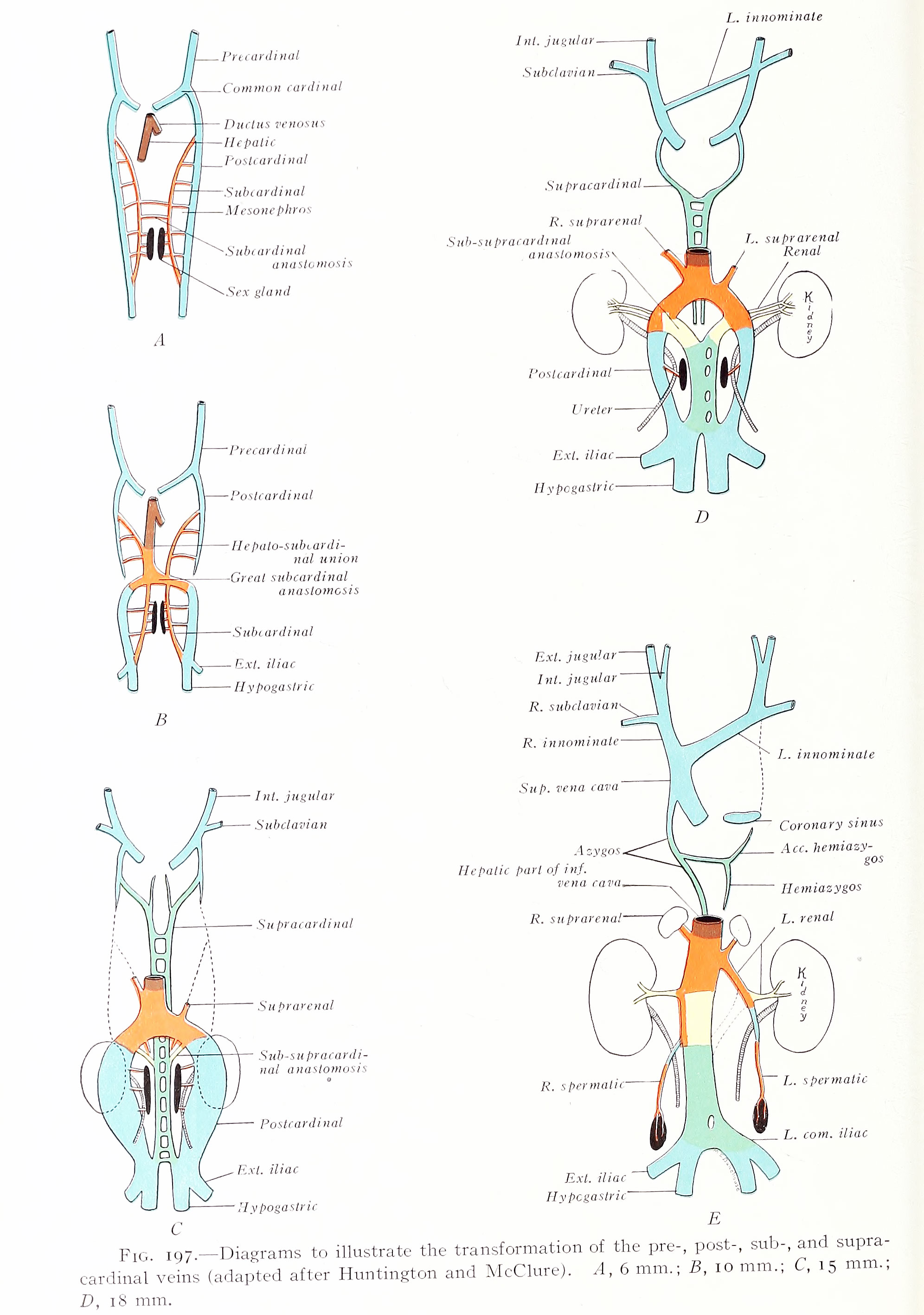 | 1,992 × 2,835 (521 KB) | Z8600021 (talk | contribs) | ==Fig. I97. Diagrams to illustrate the transformation of the pre-, post-, sub-, and supracardinal veins== (adapted after Huntington and McClure) A, 6 mm.; B, 10 mm.; , 15 mm., D, 18 mm. {{Arey1924 Footer}} Category:Cardiovascular |
You cannot overwrite this file.
File usage
The following 2 pages use this file: