File:Arey1924 fig374.jpg
From Embryology

Size of this preview: 729 × 600 pixels. Other resolution: 1,200 × 987 pixels.
Original file (1,200 × 987 pixels, file size: 268 KB, MIME type: image/jpeg)
Fig. 374. Ventral reconstruction of the cardinal and subcardinal veins in a 6 mm. pig embryo, showing the early development of the inferior vena cava ( Vehe). X 22. In the small orientation figure (cf. Fig. 376) the various jdanes are indicated by broken lines.
File history
Yi efo/eka'e gwa ebo wo le nyangagi wuncin ye kamina wunga tinya nan
| Gwalagizhi | Nyangagi | Dimensions | User | Comment | |
|---|---|---|---|---|---|
| current | 10:05, 23 October 2016 | | 1,200 × 987 (268 KB) | Z8600021 (talk | contribs) | |
| 10:04, 23 October 2016 | 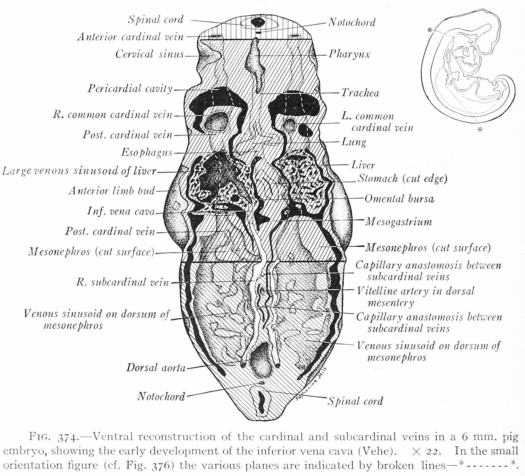 | 1,771 × 1,606 (554 KB) | Z8600021 (talk | contribs) | Fig. 374. Ventral reconstruction of the cardinal and subcardinal veins in a 6 mm. pig embryo, showing the early development of the inferior vena cava ( Vehe). X 22. In the small orientation figure (cf. Fig. 376) the various jdanes are indicated by brok... |
You cannot overwrite this file.
File usage
The following 2 pages use this file:


