File:Conklin 1905 plate06.jpg

Original file (1,521 × 2,000 pixels, file size: 680 KB, MIME type: image/jpeg)
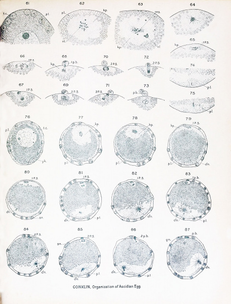
Plate VI
SeHitiits of Eggs of Cynthia partita; Maturation and Fertilization. Figs. 617S5 magnified 5S5 diameters; Figs. 70-87 magnified :.>t!<! diameters.
Fig. 61. Ovarian egg fully formed, showing germinal vesicle surrounded by yolk, and peripheral
layer of protoplasm containing test cells and yellow granules (small spheres in figure). Fig. 62. Free egg shortly after the dissolution of the nuclear membrane, showing in the middle of the
clear karyoplasm fragments of nucleolus, chromosomes and a granular mass from which
spindle fibres arise; the peripheral protoplasm contains yellow granules. Fig. 63. Egg similar to the preceding, but with the spindle fibres more fully formed. Fig. 64. Similar to preceding, spindle fibres radiate in all directions.
Fig. 65. The first polar spindle lies near the surface of the egg and its fibres are approximately paratangential ; the peripheral layer of protoplasm has streamed away from the animal pole
and the karyoplasm from the germinal vesicle has spread out here in a broad disk. Fig. 66. Metaphase of first polar spindle which is nearly parallel with surface; no centrosomes present. Fig. 67. Anaphase of first polar spindle which is turning into a radial position. Fig. OS. Separation of first polar body.
Fig. 69. Metaphase of second polar spindle, which is paratangential in position. Fig. 70. Anaphase of second polar spindle. Fig. 71. Second polar spindle approaching a. radial position. Fig. 72. Separation of second polar body.
Fig. 73. Fusion of chromosomal vesicles in egg to form egg nucleus. Fig. 74. Vegetal pole of egg of the stage shown in figs. 65 and 79, showing the entrance of the sperm
into the egg and the collection of yellow granules around the sperm head. Fig. 75. Later stage in the entrance of the sperm ; formation of sperm aster from the middle- piece. Fig. 76. Free egg before the solution of the nuclear membrane but after the extrusion of the tesl
cells; the chromosomes at the periphery of the germinal vesicle. Fig. 77. Egg after being laid but before fertilization ; chromosomes and granular substance which
forms spindle fibres in the center of the karyoplasm. The egg remains in this condition
until fertilized. Fig. 78. Same as preceding, save that spindle fibres are forming and karyoplasm has moved nearer to
the animal pole. Fig. 79. Egg showing the entrance of the spermatozoon near the vegetal pole and the spreading of
the karyoplasm into a thin cap at the animal pole. Fig. 80. Slightly more advanced stage showing development of sperm aster and collection of yellow
granules at vegetal pole, spermatozoa have entered some of the test cells. Fig. 81. First polar spindle assuming a radial position ; increase of cytoplasmic area surrounding the
sperm nucleus and aster, the latter are moving across the egg axis and hence in the
longest path toward the equator. Fig. 82. Stage slightly more advanced than the preceding; sperm nucleus, aster, clear and yellow
protoplasm becoming eccentric toward the posterior side. Fig. 83. First polar body formed ; prophase of second polar spindle. Fig. 84. Metaphase of second polar spindle; yellow protoplasm collecting into crescent. Fig. 85. Anaphase of second polar spindle, spermatozoa in some of the test cells. Fig. 86. Telophase of second polar spindle. Fig. 87. Movement of sperm nucleus and aster and of surrounding protoplasm to the posterior side
of the egg; approach of the germ nuclei.
File history
Yi efo/eka'e gwa ebo wo le nyangagi wuncin ye kamina wunga tinya nan
| Gwalagizhi | Nyangagi | Dimensions | User | Comment | |
|---|---|---|---|---|---|
| current | 15:59, 19 October 2016 |  | 1,521 × 2,000 (680 KB) | Z8600021 (talk | contribs) | ==Plate VI== SeHitiits of Eggs of Cynthia partita; Maturation and Fertilization. Figs. 617S5 magnified 5S5 diameters; Figs. 70-87 magnified :.>t!<! diameters. Fig. 61. Ovarian egg fully formed, showing germinal vesicle surrounded by yolk, and perip... |
You cannot overwrite this file.
File usage
The following 2 pages use this file: