File:Conklin 1905 plate03.jpg

Original file (1,521 × 2,000 pixels, file size: 358 KB, MIME type: image/jpeg)
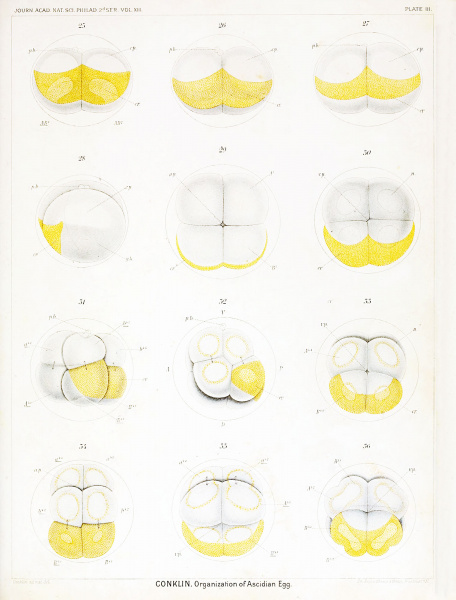
Plate III Living Eggs of Oynthia partita - First to Fourth Cleavage
Figs. 25 and 26. Same egg as the one shown in figs. 21-24; final stages in the first cleavage.
Fig. 27. Another egg at the close of the first cleavage ; seen from the posterior pole.
Fig. 28. End view of egg of same stage as preceding, showing the lateral limits of the yellow crescent, the clear protoplasm in the upper hemisphere and the yolk in the lower. The anterior portion of the lower hemisphere is composed of light gray material ; this is the gray crescent and gives rise to chorda and neural plate.
Fig. 29. Four-cell stage, viewed from the animal pole.
Fig. 30. Similar egg seen from the vegetal pole; the crescent covers about half of the posterior blastomeres.
Fig. 31. Fight-cell stage: the crescent is limited entirely to the two posterior blastomeres at the vegetal pole : while under observation the furrow between B 4' and b 4-2 shifted from the position indicated by the faint line to that shown by the heavy line, thus giving rise to the "cross furrow" shown in the next figure.
Fig. 32. Fight-cell stage, viewed from the right side, showing a small amount of yellow protoplasm around all the nuclei.
Fig. 33. Same stage viewed from the vegetal pole, showing 1% yolk laden endoderm cells and the crescent.
Fig. 34. Same stage viewed from the posterior-animal pole showing the clear ectodermal cells and the crescent.
Fig. 35. Same stage seen from the anterior-vegetal pole; yellow protoplasm around all the nuclei.
Fig. 36. Fourth cleavage of the egg seen from the vegetal pole.
File history
Yi efo/eka'e gwa ebo wo le nyangagi wuncin ye kamina wunga tinya nan
| Gwalagizhi | Nyangagi | Dimensions | User | Comment | |
|---|---|---|---|---|---|
| current | 15:32, 19 October 2016 |  | 1,521 × 2,000 (358 KB) | Z8600021 (talk | contribs) |
You cannot overwrite this file.
File usage
The following 2 pages use this file: