File:Fetal nephron development 01.jpg
Original file (1,338 × 689 pixels, file size: 119 KB, MIME type: image/jpeg)
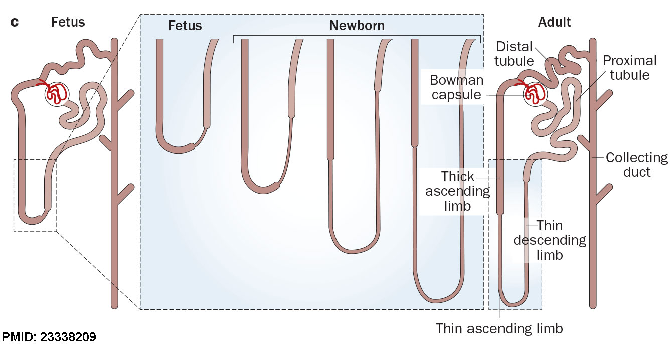
Fetal nephron development
After nephron development has completed and concomitant with the development of the renal papilla in the newborn, the thin ascending limb of Henle’s loops is generated as an outgrowth from the S3 segment of the proximal tubule and from the distal tubule anlage of the nephron. (birds and mammals)
Reference
<pubmed>23338209</pubmed>
Copyright
Reprinted by permission from Macmillan Publishers Ltd: Nature Reviews Nephrology (Romagnani, P. et al. Nat. Rev. Nephrol. 9, 137–146 (2013); published online 22 January 2013; doi:10.1038/nrneph.2012.290), copyright (2013) License Number 3954020439682
Figure 2c cropped, resized and relabelled with PMID
Cite this page: Hill, M.A. (2024, June 26) Embryology Fetal nephron development 01.jpg. Retrieved from https://embryology.med.unsw.edu.au/embryology/index.php/File:Fetal_nephron_development_01.jpg
- © Dr Mark Hill 2024, UNSW Embryology ISBN: 978 0 7334 2609 4 - UNSW CRICOS Provider Code No. 00098G
File history
Yi efo/eka'e gwa ebo wo le nyangagi wuncin ye kamina wunga tinya nan
| Gwalagizhi | Nyangagi | Dimensions | User | Comment | |
|---|---|---|---|---|---|
| current | 16:02, 22 September 2016 | | 1,338 × 689 (119 KB) | Z8600021 (talk | contribs) | |
| 16:02, 22 September 2016 |  | 1,881 × 2,293 (707 KB) | Z8600021 (talk | contribs) | ==Fetal nephron development== :'''Links''': Renal System Development ===Reference=== <pubmed>23338209</pubmed> ====Copyright==== Reprinted by permission from Macmillan Publishers Ltd: Nature Reviews Nephrology (Roma... |
You cannot overwrite this file.
File usage
The following 6 pages use this file: