File:Hydatidiform mole 02.jpg
Original file (585 × 1,100 pixels, file size: 237 KB, MIME type: image/jpeg)
Hydatidiform mole
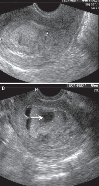
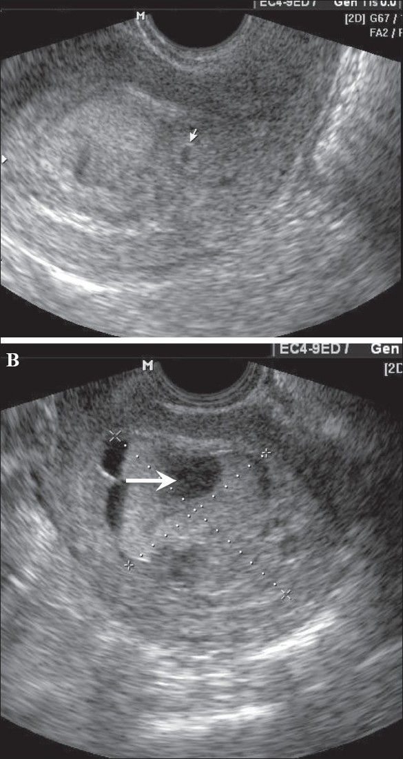
In this patient with a 6-weeks' gestation presenting with vaginal bleeding, transvaginal USG
(A) shows a gestational sac (white arrow) on the first examination. Follow-up examination after 2 days
(B) shows focal cystic changes (arrow) with loss of normal definition of the gestational sac, suggesting the possibility of molar changes. Investigations confirmed triploidy
Indian J Radiol Imaging. 2008 November; 18(4): 326–344. doi: 10.4103/0971-3026.43848.
Original file name: IJRI-18-326-g013.jpg
<pubmed>19774194</pubmed>
Copyright © Indian Journal of Radiology and Imaging
This is an open-access article distributed under the terms of the Creative Commons Attribution License, which permits unrestricted use, distribution, and reproduction in any medium, provided the original work is properly cited.
File history
Yi efo/eka'e gwa ebo wo le nyangagi wuncin ye kamina wunga tinya nan
| Gwalagizhi | Nyangagi | Dimensions | User | Comment | |
|---|---|---|---|---|---|
| current | 02:34, 27 Mayıs 2010 | | 585 × 1,100 (237 KB) | S8600021 (talk | contribs) | ==Hydatidiform mole== In this patient with a 6-weeks' gestation presenting with vaginal bleeding, transvaginal USG (A) shows a gestational sac (white arrow) on the first examination. Follow-up examination after 2 days (B) shows focal cystic changes ( |
You cannot overwrite this file.
File usage
The following page uses this file: