File:Faulconer1951 fig01.jpg
From Embryology

Size of this preview: 779 × 600 pixels. Other resolution: 1,000 × 770 pixels.
Original file (1,000 × 770 pixels, file size: 327 KB, MIME type: image/jpeg)
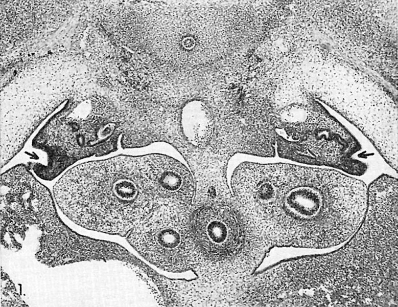
Fig. 1. Dorsolateral invagination of the ostial region of the Müllerian groove on both sides in a 13.2-mm. embryo. This location is usually described as “normal." Embryo no. 6521. X60.
File history
Yi efo/eka'e gwa ebo wo le nyangagi wuncin ye kamina wunga tinya nan
| Gwalagizhi | Nyangagi | Dimensions | User | Comment | |
|---|---|---|---|---|---|
| current | 13:54, 4 June 2016 |  | 1,000 × 770 (327 KB) | Z8600021 (talk | contribs) | |
| 13:52, 4 June 2016 |  | 1,965 × 1,231 (608 KB) | Z8600021 (talk | contribs) | Fig. 1. Dorsolateral invagination of the ostial region of the Müllerian groove on both sides in a 13.2-mm. embryo. This location is usually described as “normal." Embryo no. 6521. X60. |
You cannot overwrite this file.
File usage
The following page uses this file: