File:Gilbert1957 fig16.jpg
Original file (773 × 947 pixels, file size: 111 KB, MIME type: image/jpeg)
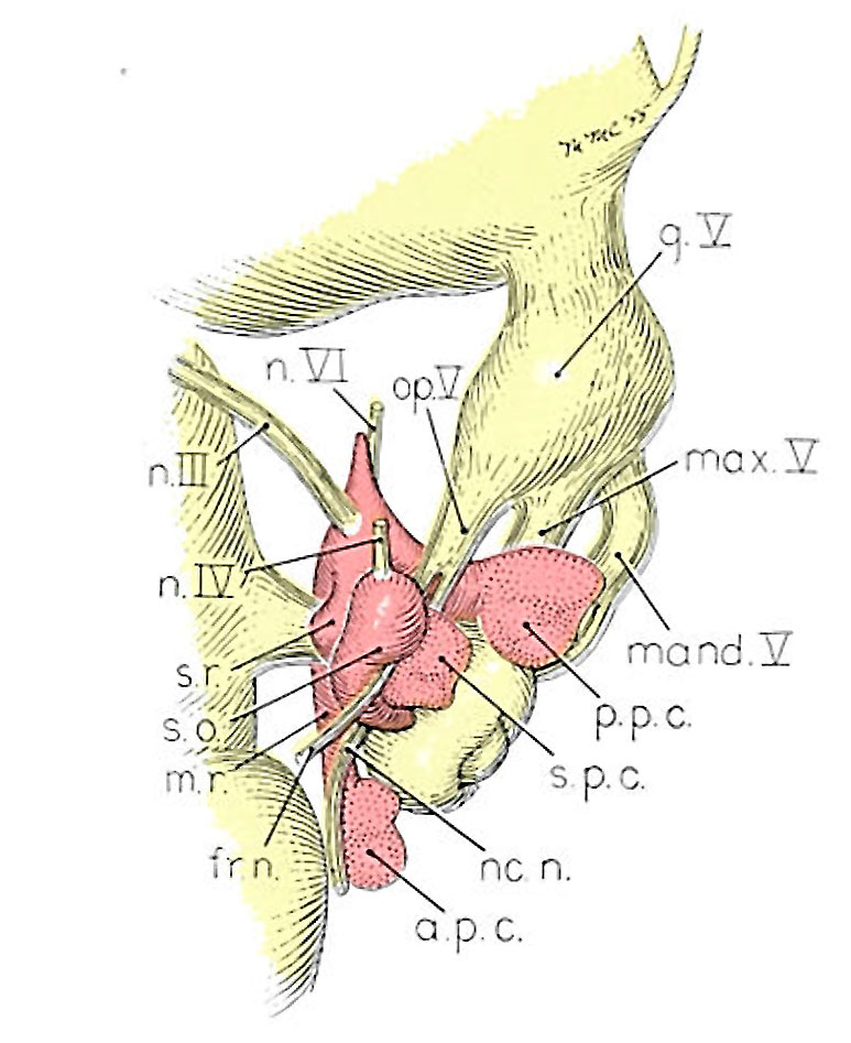
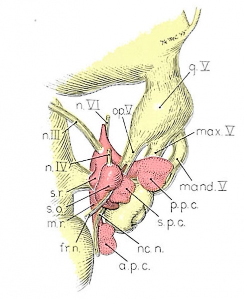
Plate 4. Four drawings of a model of the eye, eye-muscle primordia, and associated nerves,
Embryo No. 6258, horizon xvii.
Fig. 16. Dorsocranial aspect of the same model illustrated in figure 15. X30.
Attention is called to the four peripheral condensations, about the outer margin of the optic vesicle, into which the primordia of the four rectus muscles have grown. Cranial nerves III, IV, and VI have reached their respective eye-muscle primordia: the primordium of the inferior oblique has appeared as a conspicuous condensation at the distal end of the inferior rectus; a prominent bend (at the point where the trochlea will subsequently develop) has appeared near the distal end of the superior oblique primordium, and the proximal end of the superior oblique has begun to shift medially.
((Gilbert1957 figures}}
Reference
Gilbert PW. The origin and development of the human extrinsic ocular muscles. (1957) Carnegie Instn. Wash. Publ. 611, Contrib. Embryol., Carnegie Inst. Wash. 36: 59-78.
Cite this page: Hill, M.A. (2024, June 3) Embryology Gilbert1957 fig16.jpg. Retrieved from https://embryology.med.unsw.edu.au/embryology/index.php/File:Gilbert1957_fig16.jpg
- © Dr Mark Hill 2024, UNSW Embryology ISBN: 978 0 7334 2609 4 - UNSW CRICOS Provider Code No. 00098G
File history
Click on a date/time to view the file as it appeared at that time.
| Date/Time | Thumbnail | Dimensions | User | Comment | |
|---|---|---|---|---|---|
| current | 00:02, 2 June 2016 | | 773 × 947 (111 KB) | Z8600021 (talk | contribs) | ==Plate 4. Four drawings of a model of the eye, eye-muscle primordia, and associated nerves,== Embryo No. 6258, horizon xvii. Fig. 14. The eye-muscle primordia of embryo no. 6258 are superimposed on the brain of another embryo, no. 6520, of approxima... |
You cannot overwrite this file.
File usage
The following 2 pages use this file: