File:Ramsey1960-fig01.jpg
From Embryology
Size of this preview: 800 × 510 pixels. Other resolution: 1,000 × 638 pixels.
Original file (1,000 × 638 pixels, file size: 175 KB, MIME type: image/jpeg)
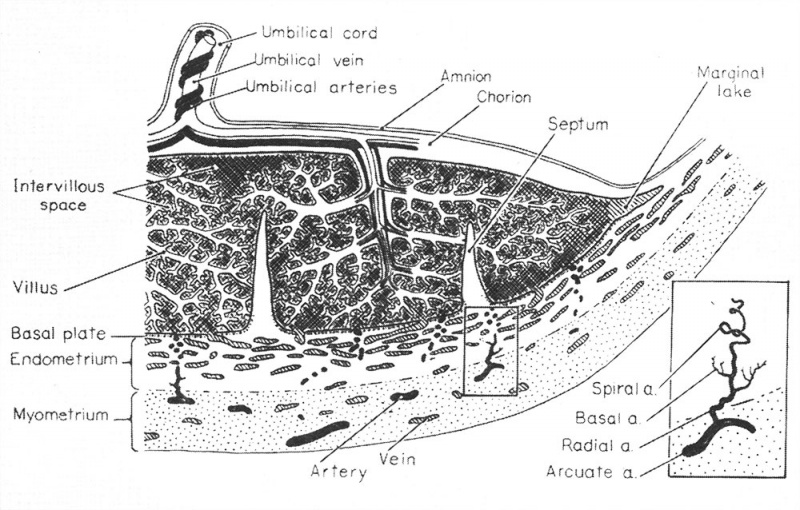
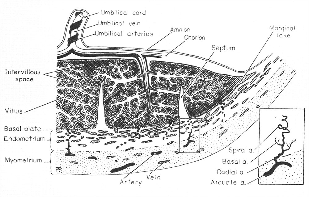
Fig. 1. Diagrammatic representation of a portion of the primate (hemochorial) placenta attached to the uterine wall
The inset shows, at higher magnification, the components of a single arterial stem.
(From Ramsey,8 in Report of 15th M&R Paediatric Research Conference 1955.)
Reference
<pubmed>16590693</pubmed>
Cite this page: Hill, M.A. (2026, October 3) Embryology Ramsey1960-fig01.jpg. Retrieved from https://embryology.med.unsw.edu.au/embryology/index.php/File:Ramsey1960-fig01.jpg
- © Dr Mark Hill 2026, UNSW Embryology ISBN: 978 0 7334 2609 4 - UNSW CRICOS Provider Code No. 00098G
File history
Yi efo/eka'e gwa ebo wo le nyangagi wuncin ye kamina wunga tinya nan
| Gwalagizhi | Nyangagi | Dimensions | User | Comment | |
|---|---|---|---|---|---|
| current | 10:40, 13 Mayıs 2016 | | 1,000 × 638 (175 KB) | Z8600021 (talk | contribs) | |
| 10:40, 13 Mayıs 2016 | 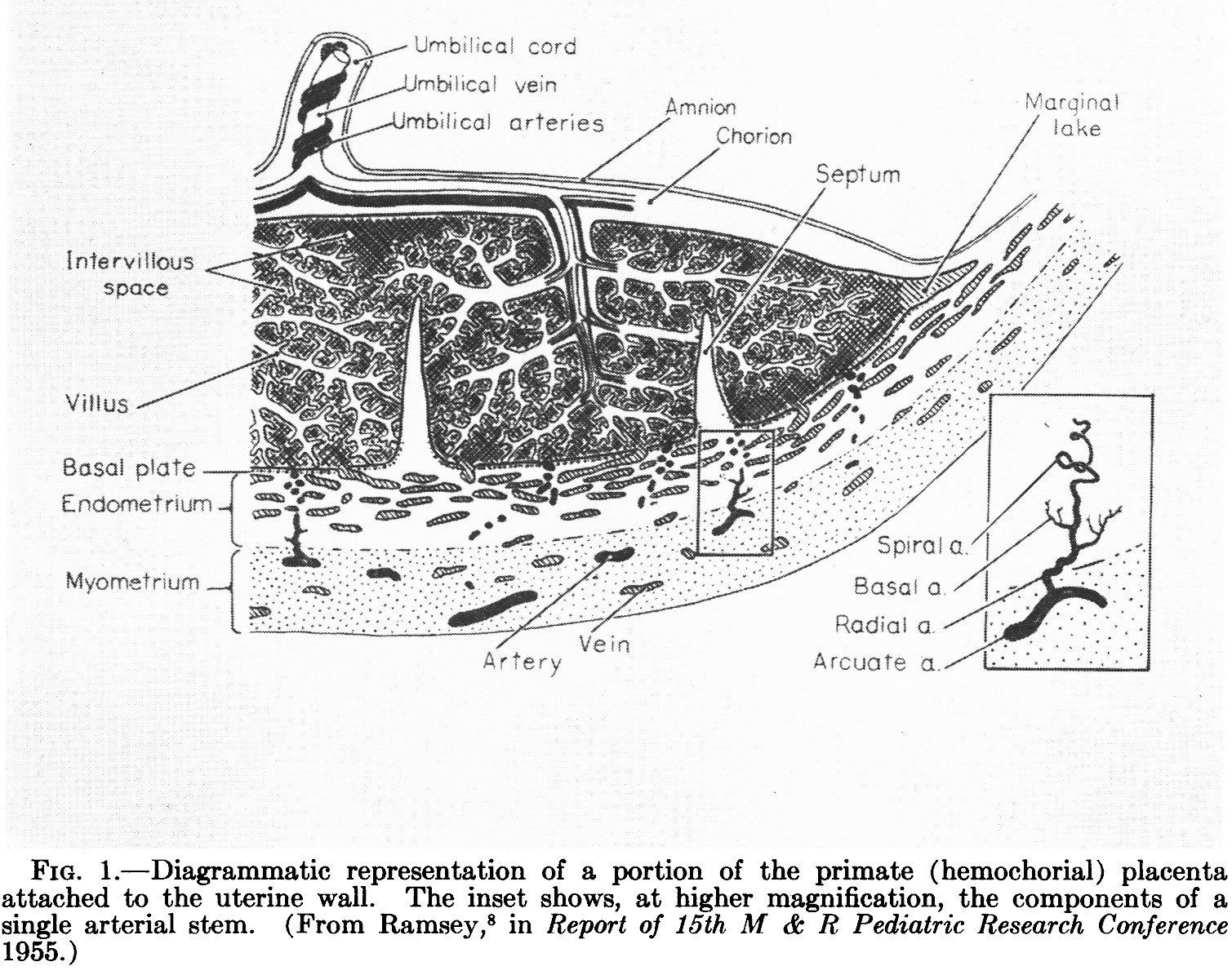 | 1,509 × 1,195 (446 KB) | Z8600021 (talk | contribs) | ==Fig. 1. Diagrammatic representation of a portion of the primate (hemochorial) placenta attached to the uterine wall== The inset shows, at higher magnification, the components of a single arterial stem. (From Ramsey,8 in Report of 15th M&R Paediatr... |
You cannot overwrite this file.
File usage
The following 3 pages use this file: