Spleen
Thymus Anatomy
| Thymus Anatomy
|
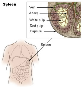
|
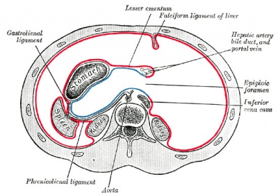
|
| left hypochondriac region
|
almost entirely surrounded by peritoneum adherent to its capsule
|
Thymus Functions
| Thymus Functions
|
- Immune - filters blood in much the way that the lymph nodes filter lymph.
- Lymphocytes in the spleen react to pathogens in the blood and attempt to destroy them.
- Macrophages then engulf the resulting debris, the damaged cells, and the other large particles.
- Red Blood Cell Removal - spleen (and liver) removes old and damaged erythrocytes from the circulating blood.
- Blood Reservoir - The sinuses in the spleen also act as a reservoir for blood. In emergencies (haemorrhage) smooth muscle in the vessel walls and in the capsule of the spleen contracts, squeezes blood out of the spleen into the general circulation.
|
Thymus Structure
| Thymus Structure
|
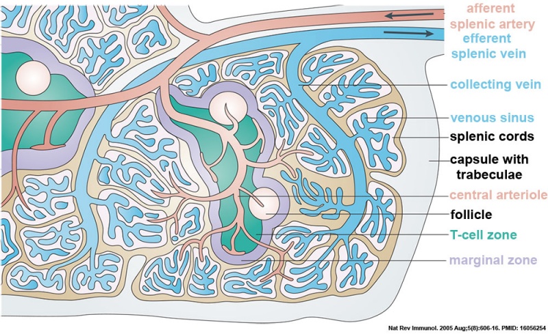
- afferent splenic artery branches into central arterioles, which are sheathed by white pulp areas.
- white pulp areas consist of the T-cell zone (also known as the periarteriolar lymphoid sheath, PALS), arterioles and B-cell follicles.
- arterioles end in cords in the red pulp, from where the blood runs into venous sinuses that collect into the efferent splenic vein.
- larger arteries and veins run together in connective-tissue trabeculae, which are continuous with the capsule that surrounds the spleen.
|
Thymus Histology
Thymus
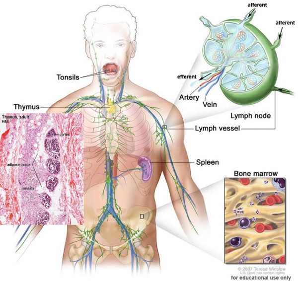
Gross Anatomy
- Superior mediastinum, anterior to heart
- Bilobed lymphoepithelial organ
Histology
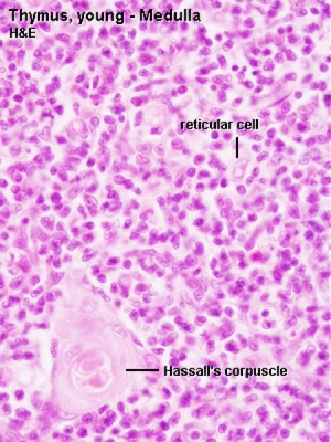
- Contains reticular cells but no fibers
- Stem lymphocytes
- proliferate and differentiate
- forms long-lived T- lymphocytes
- removes auto-reactive T-lymphocytes (negative selection)
|
|
Thymus Cells
| Reticular cells
|
Macrophages
|
Lymphocytes
|
- Abundant, eosinophilic, large, ovoid and light nucleus 1-2 nucleoli
- sheathe cortical capillaries
- form an epitheloid layer
- maintain microenvironment for development of T-lymphocytes in cortex (thymic epitheliocytes)
|
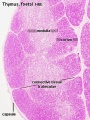
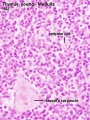
- cortex and medulla
- difficult to distinguish from reticular cells in (Stain - Haematoxylin Eosin)
- remove auto-reactive T-lymphocytes
|
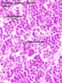
- located in cortex and medulla
- more numerous (denser) in cortex
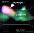
- majority are developing T-lymphocytes (= thymic lymphocytes or thymocytes)
|
Development Changes
{Changes with age
Overall Size
- birth 10-15 g
- puberty 30-40 g
- after puberty - decreases in size (involution)
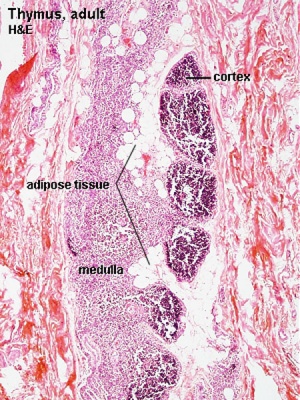
- middle-aged 10 g, replaced by adipose tissue
Histology
Adult Thymus
|
- Cortical lymphoid tissue is replaced by adipose tissue (involution)
- Increase in size of thymic corpuscles
- Thymic corpuscle - (Hassall’s corpuscle) mass of concentric epithelioreticular cells.
|