Talk:Draft 2016
From Embryology
Spleen
Spleen Anatomy
| Spleen Anatomy | |
|---|---|

|

|
| left hypochondriac region | almost entirely surrounded by peritoneum adherent to its capsule |
Spleen Function
- Immune - filters blood in much the way that the lymph nodes filter lymph.
- Lymphocytes in the spleen react to pathogens in the blood and attempt to destroy them.
- Macrophages then engulf the resulting debris, the damaged cells, and the other large particles.
- Red Blood Cell Removal - spleen (and liver) removes old and damaged erythrocytes from the circulating blood.
- Blood Reservoir - The sinuses in the spleen also act as a reservoir for blood. In emergencies (haemorrhage) smooth muscle in the vessel walls and in the capsule of the spleen contracts, squeezes blood out of the spleen into the general circulation.
Spleen Structure

- afferent splenic artery branches into central arterioles, which are sheathed by white pulp areas.
- white pulp areas consist of the T-cell zone (also known as the periarteriolar lymphoid sheath, PALS), arterioles and B-cell follicles.
- arterioles end in cords in the red pulp, from where the blood runs into venous sinuses that collect into the efferent splenic vein.
- larger arteries and veins run together in connective-tissue trabeculae, which are continuous with the capsule that surrounds the spleen.
- Capsule, trabeculae (dense connective tissue)
- Splenic pulp white pulp, red pulp - based on appearance and cell content.

|
White Pulp
|
Red Pulp
|
Reticular Fibers (type III collagen) act as supporting meshwork.
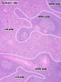
Overview Red and White Pulp
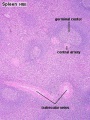
Overview Red and White Pulp
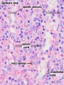
Cords and Sinuses
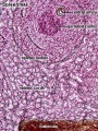
Reticular Fibre overview
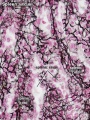
Reticular Fibre detail
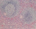
unlabeled red and white pulp
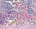
unlabeled red pulp and macrophages
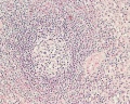
unlabeled white pulp germinal centre
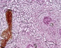
unlabeled reticular fibre
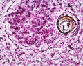
unlabeled white pulp reticular
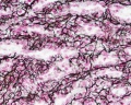
unlabeled red pulp reticular


