Ovary Development

Introduction

The female gonad is the ovary and is closely associated with female internal genital (reproductive) tract development. In humans, these laterally paired organs lie within the peritoneal cavity. Genes such as Wnt-4 and DAX-1 necessary for initiation of female pathway ovary development, female gonad is not considered a default process.
Initial gonad development in females and males is virtually identical with germ cells migrating into an indifferent gonad. In females with XX, the ovary then begins to develop and the subsequent structure and timecourse of germ cell then differs between males and females. In the ovary oocytes proliferate prior to birth and arrest in meiosis 1.
| Menstrual Cycle | X Chromosome | Genital System - Female | Oogenesis
Human Timeline
Approximate Timeline of human development listed below.
24 days - intermediate mesoderm, pronephros primordium
28 days - mesonephros and mesonephric duct
35 days - uteric bud, metanephros, urogenital ridge
42 days - cloacal divison, gonadal primordium (indifferent)
49 days - paramesonephric duct, gonadal differentiation
56 days - paramesonephric duct fusion (female)
100 days - primary follicles (ovary)
Oogenesis

The 2 human ovaries gradually lose follicles both before and after puberty (the beginning of ovulation); beginning with about 7 million before birth, 2 million at birth, 300-400,000 by puberty and finally by late 40’s have only a few follicles left. The number of antral follicles detected within the ovary also decreases with increasing materal age. In humans, a primodial follicle take about 150 days to develop into a preantral follicle (primary) and another 120 days to form an antral follicle (secondary). A number of antral follicles will then "compete" for 14-15 days to become the dominant follicle, which will undergo ovulation.
- The graph shows the changes in human germ cell numbers in the ovary with age.
- Total numbers peak at about 7 million (occuring in early fetal development) and then decreasing by apopotic cell death.
- At puberty there remain only about 400,000 and only about 10% of these will be released through reproductive life.
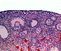
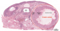
Infant Ovary

This image shows a region (see inset) of the infant ovary cortex.
There are a large number of developing oocytes which will eventually form a dense primordial germ layer at the ovary periphery.
Later stages of follicle development are completely absent and will begin to only appear just prior to puberty.
Postnatal Oogenesis
There is a dogma in mammalian development that new oocyte and follicle production does not occur during postnatal life. There is substantial data that shows human ovarian changes postnatally are loss by apoptosis of prenatal oocytes. A research group (Tilly JL, Johnson J. 2004, 2007) has recently published experiments using mice, showing potentially other sources/sites (bone marrow) of oocyte (putative germ cell) generation. They recently stated that the argument should be based upon "experimental approaches than simply an absence of evidence, especially from gene expression analyses". Several other research groups (Eggan K etal. 2004 and Veitia etal. 2007) have argued against these findings.
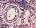
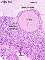
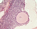
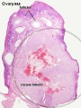
Adult Follicle Structure

A follicle usually contains a single oocyte (egg, ovum, female gamete) and a series of supporting cells and a single fluid-filled space in layers surrounding this cell. The 3 layers below are arranged in layers outward from the oocyte.
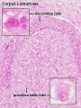
Granulosa Cells
- A specific cell type that proliferates in association with the oocyte within the developing follicles of the ovary. These cells form the follicle stratum granulosa and are also given specific names based upon their position within the follicle.
- The membrana granulosa cells sit on the follicular basal lamina and line the antrum as a stratified epitelium. Following ovulation, these granulosa cells contribute to corpus luteum.
- The cumulus oophorus is a column of granulosa cells that attaches the oocyte to the follicle wall. At ovulation, this column of cells is broken or separates to release the oocyte from its follicle attachment.
- The corona radiata are the granulosa cells that directly surround the oocyte, and are released along with it at ovulation. Following ovulation, the corona radiata provide physical protection to the oocyte and are the initial structural barrier that spermatazoa must penetrate during fertilization.
Follicular Fluid
- The antrum is a fluid-filled space in the secondary (antral) follicle
- At ovulation, fluid is released along with the oocyte
- Thought to "carry" the oocyte out of the follicle (like a boat on a wave)
- Aids entry into the uterine tube
Theca Interna
(Greek, thek = box) The ovarian follicle endocrine cells forming the inner layer of the theca folliculi surrounding the developing follicle within the ovary. This vascularized layer of cells respond to leutenizing hormone (LH) synthesizing and secreting androgens (androstendione) transported to glomerulosa cells which process initially into testosterone and then by aromatase into estrogen (estradiol). Theca cells do not begin hormonal functions until puberty.
Theca Externa
(Greek, thek = box) The ovarian follicle stromal cells forming the outer layer of the theca folliculi surrounding the developing follicle within the ovary. Consisting of connective tissue cells, smooth muscle and collagen fibers.
Follicle Factors
There are both external endocrine factors and follicle internal factors that can influence the development and atresia of ovarian follicles.
External Factors
- Leutenizing Hormone (LH)
- from the anterior pituitary
- stimulate the theca interna to synthesize and secrete androgens (androstendione) transported to glomerulosa cells
- glomerulosa cells process initially into testosterone and then by aromatase into estrogen (estradiol)
- Follicle-stimulating hormone (FSH)
- from the anterior pituitary
- initiates follicule growth through the granulosa cells
- involved in selecting the most advanced (sensitive) follicle to proceed to ovulation
Internal Factors
- Oocyte Factors
- growth differentiation factor-9 (GDF-9) - involved in the differentiation of theca cells during this early stage of follicular development
- Granulosal Factor(s)
- stimulates the recruitment of theca cells from cortical stromal cells
- Thecal Factor(s)
- Epidermal growth factor (EGF), TGF-α, keratinocyte growth factor (KGF), hepatocyte growth factor (HGF), and BMP-7 - appear to be several inhibitors of apoptotic cell death
Follicle Classification
There are several different nomenclatures for the stages of follicle maturation. It probably does not matter which naming system you use, as long as you are consistent and use the same set of terminology for all stages.

Primordial Follicle
Alternate nomenclature: small follicle or type 1, 2, 3 (25cells)
Primary Follicle
Alternate nomenclature: preantral follicle or type 4 (26-100 cells), type 5 (101-300 cells)
Secondary Follicle
Alternate nomenclature: small and large antral follicle or type 6 (3001-500 cells), type 7 (501-1000 cells)
Preovulatory Follicle
Alternate nomenclature: Graafian follicle or type 8 (>1000 cells)
Atresia
At any one time the majority of follicles are destined not to complete maturation and at any stage (from type 4-7) degeneration of the follicle can occur. This process is called ATRESIA.
References
Reviews
Articles
- Oocyte-granulosa-theca cell interactions during preantral follicular development. Orisaka M, Tajima K, Tsang BK, Kotsuji F. J Ovarian Res. 2009 Jul 9;2(1):9. PMID: 19589134 | J Ovarian Res.
- The forkhead transcription factor FOXL2 is expressed in somatic cells of the human ovary prior to follicle formation. Duffin K, Bayne RA, Childs AJ, Collins C, Anderson RA. Mol Hum Reprod. 2009 Dec;15(12):771-7. Epub 2009 Aug 25. PMID: 19706741 | Mol Hum Reprod.
Search Pubmed
Search Pubmed: Ovary Development | Follicle Development | Follicle Atresia
Additional Images
Cat ovary histology
ovary-primary follicle, primordial follicle, oocyte histology

Primary follicle histology
Secondary follicle histology
- Monkey-ovary x20he.jpg
Monkey ovary
Secondary Follicle
Corpus luteum histology
Corpus luteum histology
Corpus luteum lutein cells histology
Ovary- follicle stages
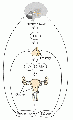
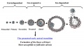
The female hypothalamic–pituitary–gonadal axis
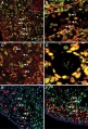
Human fetal ovary - FOXL2 expression


