File:Fetal adrenal gland steroidogenesis.jpg

Fetal_adrenal_gland_steroidogenesis.jpg (778 × 600 pixels, file size: 80 KB, MIME type: image/jpeg)
Fetal Adrenal Gland Steroidogenesis
| Ontogenesis of steroidogenic enzymes in the human fetal adrenal gland. This schematic representation is divided into portions showing the fetal adrenal gland (right) at the first, second and third trimesters of pregnancy, and the adult adrenal gland (left). During the first trimester, the fetal gland is composed of a definitive zone (DZ, light grey) and a fetal zone (FZ, dark grey).
The fetal zone, expressing the P450C17 cytochrome, is responsible for massive secretion of DHEA and DHEA/S, used by the placenta as estrogen precursors. During the 2nd trimester, chromaffin cells (CC, black) originating from the neural crests migrate through the fetal cortex to progressively colonize the center of the gland to form the future medulla (Med). At the end of the 2nd trimester, the newly constituted transitional zone (TZ, medium grey) acquires the enzyme 3ß-HSD while the expression of P450C17 remains, thus allowing the production of fetal cortisol. Near birth, cells of the definitive zone which express only 3ß-HSD, acquire the P450aldo and begin to secrete mineralocorticoids such as aldosterone. Shortly after birth, the fetal adrenal regresses strongly (mainly due to the regression of the fetal zone) and recovers progressively during the first years of extra-uterine life. Finally, the adult adrenal gland is composed of the zona glomerulosa (ZGlo, light grey), zona fasciculata (ZFasc , medium grey) and zona reticularis (ZRet, dark grey) responsible for the production of mineralocorticoids (aldosterone), glucocorticoids (cortisol) and androgens (DHEA-DHEA/S), respectively. |
|
- Links: Adrenal Development
Reference
<pubmed>16172742</pubmed>| Braz J Med Biol Res.
Copyright
All the contents of www.scielo.br, except where otherwise noted, is licensed under a Creative Commons Attribution License.
Figure 1. 5928i01.jpg http://www.scielo.br/img/revistas/bjmbr/v38n10//html/5928i01.htm
Cite this page: Hill, M.A. (2024, June 16) Embryology Fetal adrenal gland steroidogenesis.jpg. Retrieved from https://embryology.med.unsw.edu.au/embryology/index.php/File:Fetal_adrenal_gland_steroidogenesis.jpg
- © Dr Mark Hill 2024, UNSW Embryology ISBN: 978 0 7334 2609 4 - UNSW CRICOS Provider Code No. 00098G
File history
Click on a date/time to view the file as it appeared at that time.
| Date/Time | Thumbnail | Dimensions | User | Comment | |
|---|---|---|---|---|---|
| current | 08:33, 11 November 2015 | | 778 × 600 (80 KB) | Z8600021 (talk | contribs) | |
| 08:24, 11 November 2015 | 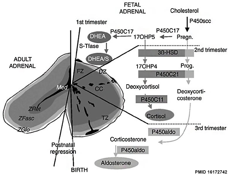 | 778 × 600 (71 KB) | Z8600021 (talk | contribs) | Figure 1. Ontogenesis of steroidogenic enzymes in the human fetal adrenal gland. This schematic representation is divided into portions showing the fetal adrenal gland (right) at the first, second and third trimesters of pregnancy, and the adult adrena... |
You cannot overwrite this file.
File usage
The following 4 pages use this file: