File:Morton1949 plate02.jpg
From Embryology
Size of this preview: 383 × 599 pixels. Other resolution: 1,598 × 2,499 pixels.
Original file (1,598 × 2,499 pixels, file size: 1 MB, MIME type: image/jpeg)
Plate 1
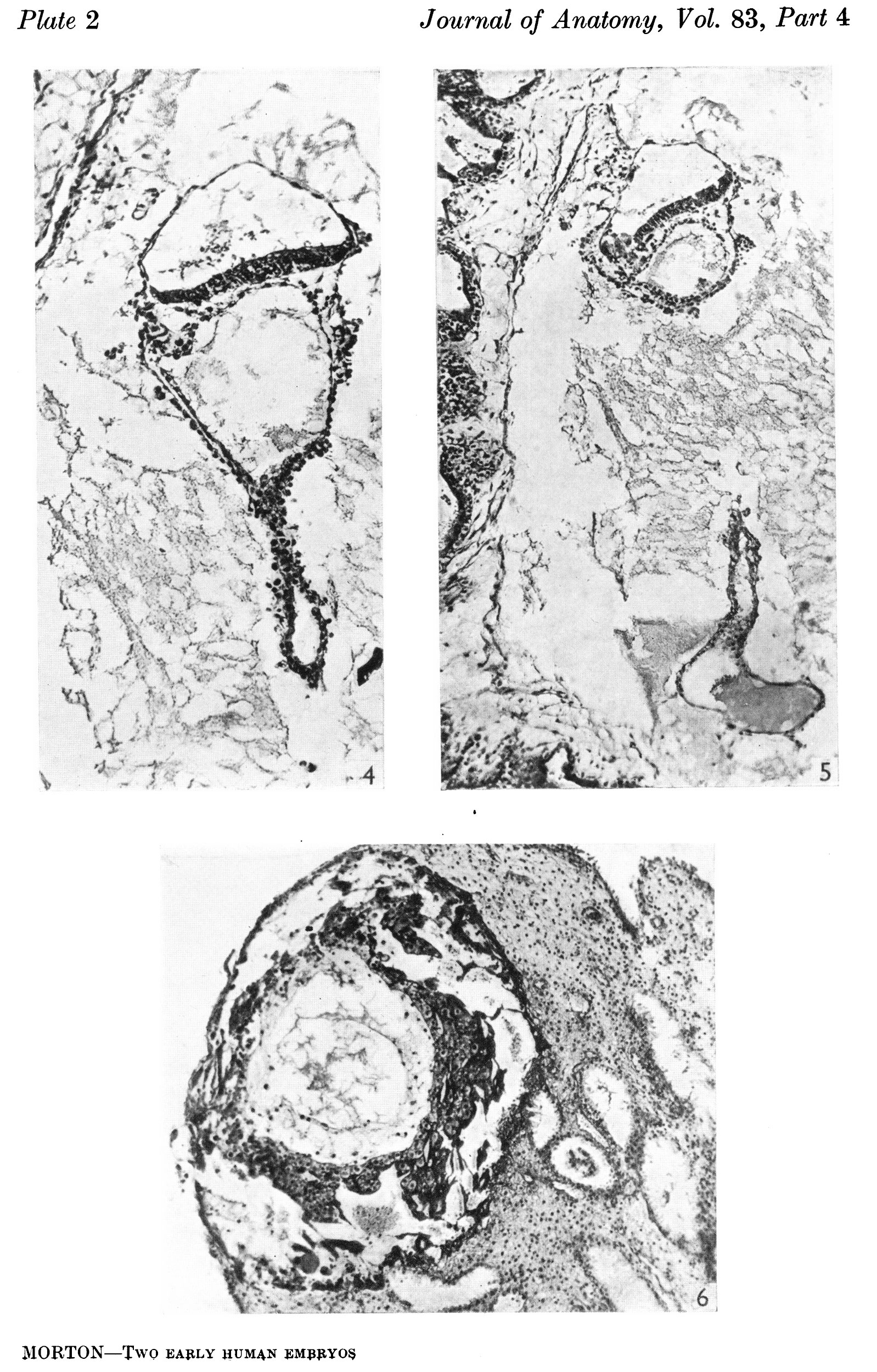
Fig. 1. The ovum in 8itu. Sect. 81. x 38.
Fig. 2. The duct part of the yolk-sac. The distal end is inferior. Sect. 77. x 375.
Fig. 3. The distal expansion of the yolk-sac. The end of the duct just appears above, on the left. Sect.77. x375.
File history
Yi efo/eka'e gwa ebo wo le nyangagi wuncin ye kamina wunga tinya nan
| Gwalagizhi | Nyangagi | Dimensions | User | Comment | |
|---|---|---|---|---|---|
| current | 21:09, 9 August 2015 | | 1,598 × 2,499 (1 MB) | Z8600021 (talk | contribs) | ==Plate 1== Fig. 1. The ovum in 8itu. Sect. 81. x 38. Fig. 2. The duct part of the yolk-sac. The distal end is inferior. Sect. 77. x 375. Fig. 3. The distal expansion of the yolk-sac. The end of the duct just appears above, on the left. Sect.77. x375. |
You cannot overwrite this file.
File usage
The following page uses this file:


