File:Streeter1906 fig03.jpg
Original file (1,472 × 1,943 pixels, file size: 285 KB, MIME type: image/jpeg)
FIG.4. Diagram representing the growth and stages of differentiation of the human membranous labyrinth. At 31h weeks (6-7 mm.) the ear vesicle consists of two simple pouches, into the upper of which opens the endolymphatic appendage. At 4 weeks sac.endolymph. ,sac, endolymph. duct. endolymph., (9 mm.) there is at the base of the vestibular pouch an atrium, the space destined to form the utricle and saccule. At 5 weeks (12 mm.) this space is circumscribed from the cochlear pouah below by a constriction corresponding to the ductus reuniens, and above Prom the rest of the vestibular pouch by the formation of the semicircular canals. At 6 weeks (20 mm.) an ingrowth of the wall of the atrium divides it intoanupper part (utricle) and lower part (saccule). At 10 weeks (30 mm.) this partition between the utricle and saccule is complete and extends inward .in such a way asto split the orifice of the endolymphatic duct.
| Historic Disclaimer - information about historic embryology pages |
|---|
 |
- Mall 1906 Links: Fig 1. 14mm Embryo | Fig 2. 30mm Embryo | Fig 3. Semicircular canal | Fig 4. Membranous Labyrinth | Fig 5. Acoustic nerve complex | Fig 6. Facial-acoustic Complex | Fig 7. Facial Nerve Pig Embryo 20 cm | Fig 8. Geniculate Ganglion | Plate 1. Human Embryo 4 to 20 mm | Plate 2. Human Embryo 30 mm | Membranous Labyrinth and Nerves
Fig 1 Membranous Labyrinth Human Embryo 14 mm
Fig 2 30mm Embryo
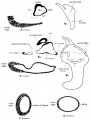
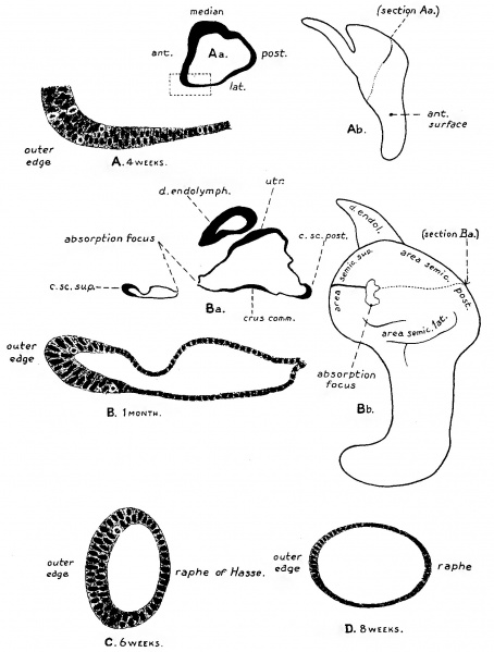
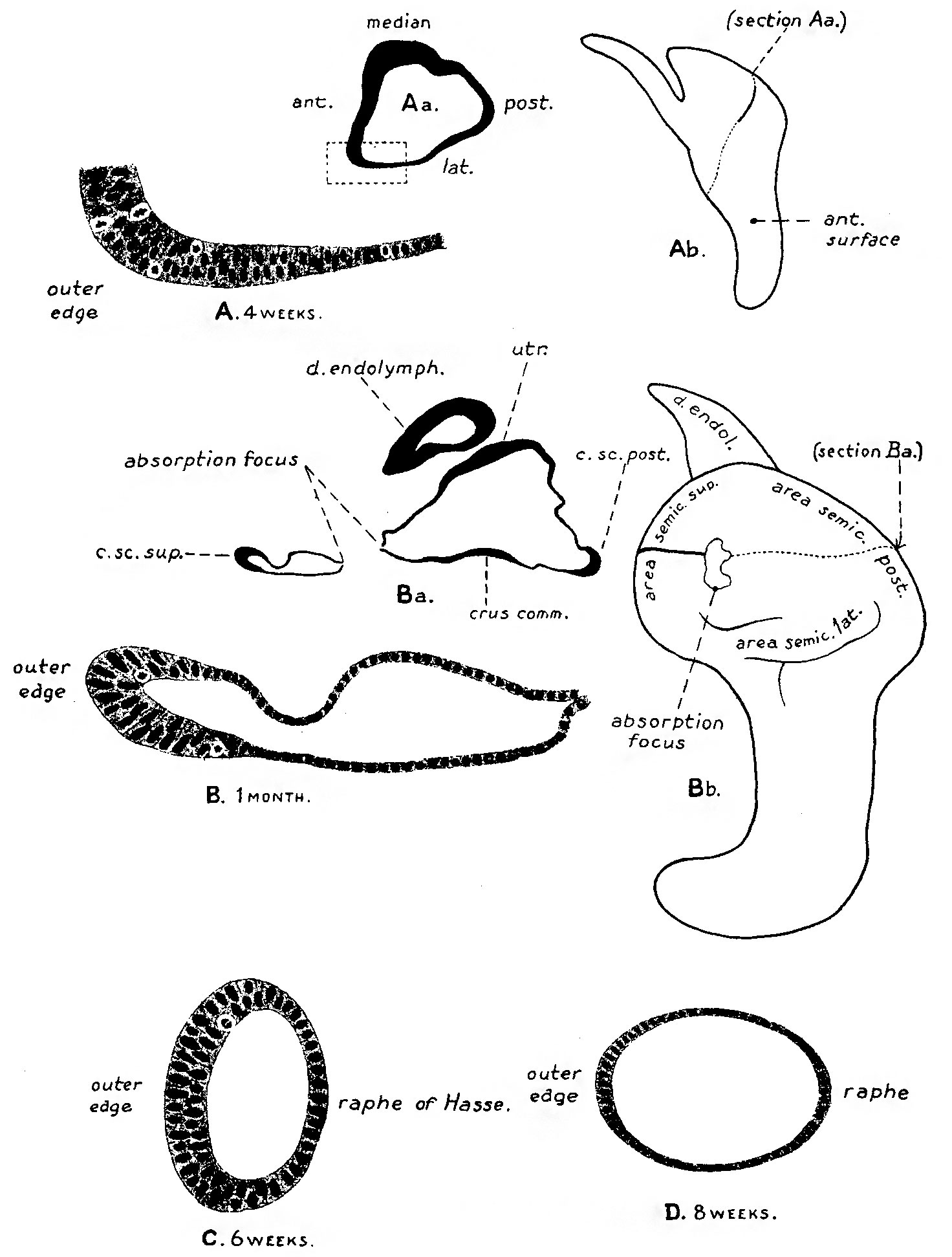
Fig 3 Semicircular canal
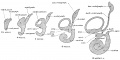
Fig 4 Membranous Labyrinth Growth
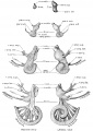
Fig 5 Acoustic nerve complex
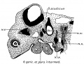
Fig 6 Facial-acoustic Complex Human Embryo 7 mm
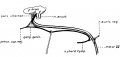
Fig 7 Facial Nerve Pig Embryo 20 cm
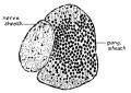
Fig 8 Geniculate Ganglion Human Embryo 30 mm
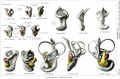
Plate 1. Membranous Labyrinth Human Embryo 4 to 20 mm
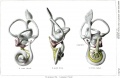
Plate 2. Membranous Labyrinth Human Embryo 30 mm


Reference
Streeter GL. On the development of the membranous labyrinth and the acoustic and facial nerves in the human embryo. (1906) Amer. J Anat. 6:139-165.
Cite this page: Hill, M.A. (2026, October 4) Embryology Streeter1906 fig03.jpg. Retrieved from https://embryology.med.unsw.edu.au/embryology/index.php/File:Streeter1906_fig03.jpg
- © Dr Mark Hill 2026, UNSW Embryology ISBN: 978 0 7334 2609 4 - UNSW CRICOS Provider Code No. 00098G
File history
Yi efo/eka'e gwa ebo wo le nyangagi wuncin ye kamina wunga tinya nan
| Gwalagizhi | Nyangagi | Dimensions | User | Comment | |
|---|---|---|---|---|---|
| current | 18:10, 23 July 2015 | | 1,472 × 1,943 (285 KB) | Z8600021 (talk | contribs) | |
| 18:10, 23 July 2015 |  | 1,472 × 2,200 (384 KB) | Z8600021 (talk | contribs) |
You cannot overwrite this file.
File usage
The following 14 pages use this file:
- Carnegie stage 23
- Hearing - Inner Ear Development
- Paper - On the development of the membranous labyrinth and the acoustic and facial nerves in the human embryo
- File:Streeter1906 fig01.jpg
- File:Streeter1906 fig02.jpg
- File:Streeter1906 fig03.jpg
- File:Streeter1906 fig04.jpg
- File:Streeter1906 fig05.jpg
- File:Streeter1906 fig06.jpg
- File:Streeter1906 fig07.jpg
- File:Streeter1906 fig08.jpg
- File:Streeter1906 plate01.jpg
- File:Streeter1906 plate02.jpg
- Template:Streeter1906 figures