File:Anson1934 fig01-8.jpg
From Embryology
Size of this preview: 800 × 531 pixels. Other resolution: 1,337 × 888 pixels.
Original file (1,337 × 888 pixels, file size: 126 KB, MIME type: image/jpeg)
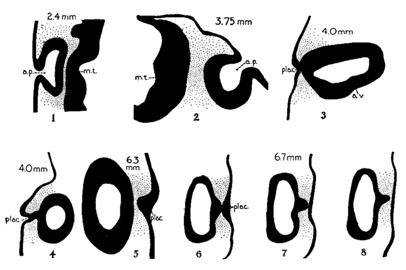
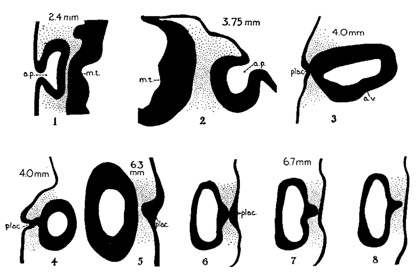
Figs. 1 to 8. Auditory Region - Transverse sections of embryos in the Harvard Embryological Collection
Dorsal aspect downward (except in 3 ) . x 80.
|
Abbreviations
a.p., auditory pit a.v., auditory vesicle (otocyst) c.d., cochlear duct coch., cochlear part of vesicle e.ap., endolymphatic appendage e.d., endolymphatic duct e.pr., eiidolymphatic projeetion e.s., endolymphatic sac ep., epidermis l.s.d., lateral semicircular duct m.t., medullary tube plac., placodal stalk sacc., saccule s.s.d., superior semicircular duct utr., utricle vest., vesibular part of vesicle |
File history
Yi efo/eka'e gwa ebo wo le nyangagi wuncin ye kamina wunga tinya nan
| Gwalagizhi | Nyangagi | Dimensions | User | Comment | |
|---|---|---|---|---|---|
| current | 14:52, 23 July 2015 | | 1,337 × 888 (126 KB) | Z8600021 (talk | contribs) | |
| 14:50, 23 July 2015 |  | 1,800 × 1,556 (384 KB) | Z8600021 (talk | contribs) |
You cannot overwrite this file.
File usage
The following 2 pages use this file: