File:Hypospadia 3D ultrasound 01.jpg
From Embryology

Size of this preview: 800 × 346 pixels. Other resolution: 1,150 × 497 pixels.
Original file (1,150 × 497 pixels, file size: 87 KB, MIME type: image/jpeg)
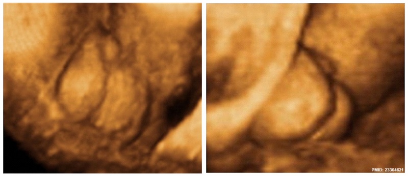
Penoscrotal Hypospadia 3D Ultrasound
Ultrasonography in rendering mode, at ((GA)) 33 weeks, with short penis and with evidence of testicles inside a bifid scrotum.
Figure 2 http://www.hindawi.com/journals/criu/2012/142814/fig2/
File history
Yi efo/eka'e gwa ebo wo le nyangagi wuncin ye kamina wunga tinya nan
| Gwalagizhi | Nyangagi | Dimensions | User | Comment | |
|---|---|---|---|---|---|
| current | 07:54, 17 May 2015 | | 1,150 × 497 (87 KB) | Z8600021 (talk | contribs) | ==Penoscrotal Hypospadia 3D Ultrasound== Ultrasonography in rendering mode, at ((GA)) 33 weeks, with short penis and with evidence of testicles inside a bifid scrotum. Figure 2 http://www.hindawi.com/journals/criu/2012/142814/fig2/ |
You cannot overwrite this file.
File usage
The following 2 pages use this file: