File:Fetal cardiac ultrasound 01.jpg
Original file (900 × 600 pixels, file size: 88 KB, MIME type: image/jpeg)
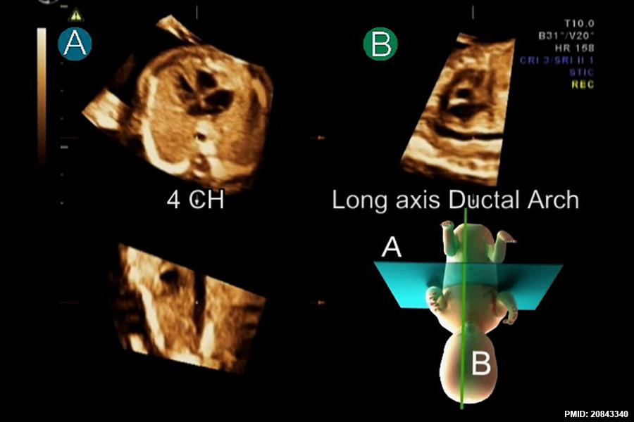
After standardization of fetal position in the STIC volume dataset to place the fetus in the exact dorsal supine position, navigating systematically in the volume usually provides a reproducible image from a corresponded movement. Placing the reference dot in the center of the aorta in the four-chamber (4 CH) view in plane A simultaneously displays long axis of the ductal arch in plane B.
Jantarasaengaram and Vairojanavong Cardiovascular Ultrasound 2010 8:41
1476-7120-8-41-3.jpg
doi:10.1186/1476-7120-8-41
Cardiovasc Ultrasound. 2010 Sep 15;8:41. doi: 10.1186/1476-7120-8-41. Eleven fetal echocardiographic planes using 4-dimensional ultrasound with spatio-temporal image correlation (STIC): a logical approach to fetal heart volume analysis. Jantarasaengaram S1, Vairojanavong K.
Abstract
BACKGROUND: Theoretically, a cross-sectional image of any cardiac planes can be obtained from a STIC fetal heart volume dataset. We described a method to display 11 fetal echocardiographic planes from STIC volumes. METHODS: Fetal heart volume datasets were acquired by transverse acquisition from 200 normal fetuses at 15 to 40 weeks of gestation. Analysis of the volume datasets using the described technique to display 11 echocardiographic planes in the multiplanar display mode were performed offline. RESULTS: Volume datasets from 18 fetuses were excluded due to poor image resolution. The mean visualization rates for all echocardiographic planes at 15-17, 18-22, 23-27, 28-32 and 33-40 weeks of gestation fetuses were 85.6% (range 45.2-96.8%, N = 31), 92.9% (range 64.0-100%, N = 64), 93.4% (range 51.4-100%, N = 37), 88.7%(range 54.5-100%, N = 33) and 81.8% (range 23.5-100%, N = 17) respectively. CONCLUSIONS: Overall, the applied technique can favorably display the pertinent echocardiographic planes. Description of the presented method provides a logical approach to explore the fetal heart volumes.
PMID 20843340
File history
Yi efo/eka'e gwa ebo wo le nyangagi wuncin ye kamina wunga tinya nan
| Gwalagizhi | Nyangagi | Dimensions | User | Comment | |
|---|---|---|---|---|---|
| current | 12:53, 10 Mayıs 2015 | | 900 × 600 (88 KB) | Z8600021 (talk | contribs) | After standardization of fetal position in the STIC volume dataset to place the fetus in the exact dorsal supine position, navigating systematically in the volume usually provides a reproducible image from a corresponded movement. Placing the reference... |
You cannot overwrite this file.
File usage
There are no pages that use this file.


