File:Spaulding-fig01.jpg
Original file (934 × 743 pixels, file size: 101 KB, MIME type: image/jpeg)
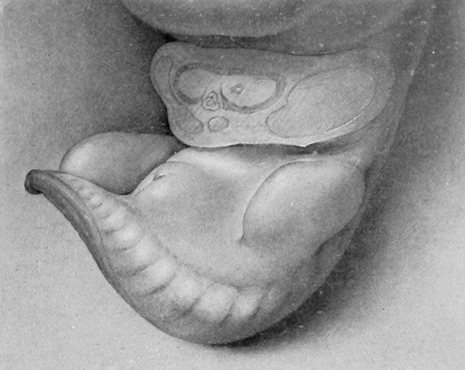
Fig. 1. Carnegie Embryo No. 467
9 mm X 21.
Stage 1, 9 mm. (fig. 1, plate 1). The earliest stage that I have studied is shown in an embryo 9 mm. long. In this specimen the genital tubercle is a very low conical eminence between the umbilical cord and the base of the tail. Its apex is separated from the rest of the tubercle by a pair of converging depressions which unite in the midventral line to form a longitudinal groove extending to the caudal border, giving to the entire groove a Y-shape. Caudal to the arms of the Y and lateral to its stem is a pair of broad, swollen areas. This condition is slightly different from the stage described by Keibel (1896) of the beginning development of the genitalia in a 3-mm. embryo. In his specimen he described the cloacal tubercle as consisting of a pair of eminences separated by the cloacal membrane. The difference between the stage described by Keibel and my first stage may be accounted for entirely by the difference in the size of the two specimens.
| Historic Disclaimer - information about historic embryology pages |
|---|
 |
- Figure Links: Text | Text Figure 1 | Text Figure 2 | Plate 1 | Fig. 1 | Fig. 2 | Fig. 3 | Fig. 4 | Fig. 5 | Fig. 6 | Plate 2 | Fig. 7 | Fig. 8 | Fig. 9 | Fig. 10 | Fig. 11 | Fig. 12 | Fig. 13 | Fig. 14 | Fig. 15 | Fig. 16 | Fig. 17 | Fig. 18 | Fig. 19 | Fig. 20 | Fig. 21 | Fig. 22 | Plate 3 | Fig. 23 | Fig. 24 | Fig. 25 | Fig. 26 | Fig. 27 | Fig. 28 | Fig. 29 | Plate 4 | Fig. 30 | Fig. 31 | Fig. 32 |Fig. 33 | Fig. 34 | Fig. 35 | Fig. 36 | Fig. 37 | Fig. 38 | Fig. 39 | Fig. 40 | Fig. 41 | Fig. 42 | Fig. 43 | Fig. 44 | Fig. 45 | Fig. 46 | Fig. 47 | Fig. 48 | Fig. 49 | Fig. 50 | Fig. 51 | Fig. 52 | Fig. 53 | Fig. 54
| Historic Disclaimer - information about historic embryology pages |
|---|
|
Reference
Spaulding MH. The development of the external genitalia in the human embryo. (1921) Contrib. Embryol., Carnegie Inst. Wash. Publ. 81, 13: 69 – 88.
Cite this page: Hill, M.A. (2024, June 15) Embryology Spaulding-fig01.jpg. Retrieved from https://embryology.med.unsw.edu.au/embryology/index.php/File:Spaulding-fig01.jpg
- © Dr Mark Hill 2024, UNSW Embryology ISBN: 978 0 7334 2609 4 - UNSW CRICOS Provider Code No. 00098G
Reference
Spaulding, M.H., The development of the external genitalia in the human embryo. Contributions to Embryology Carnegie Institution No.61 (1921). With four plates and two text-figures.
Cite this page: Hill, M.A. (2024, June 15) Embryology Spaulding-fig01.jpg. Retrieved from https://embryology.med.unsw.edu.au/embryology/index.php/File:Spaulding-fig01.jpg
- © Dr Mark Hill 2024, UNSW Embryology ISBN: 978 0 7334 2609 4 - UNSW CRICOS Provider Code No. 00098G
File history
Click on a date/time to view the file as it appeared at that time.
| Date/Time | Thumbnail | Dimensions | User | Comment | |
|---|---|---|---|---|---|
| current | 23:22, 14 April 2015 | | 934 × 743 (101 KB) | Z8600021 (talk | contribs) |
You cannot overwrite this file.
File usage
The following 2 pages use this file: