File:Sinus venosus atrial septal defect 01.jpg
Original file (1,000 × 754 pixels, file size: 90 KB, MIME type: image/jpeg)
24987269
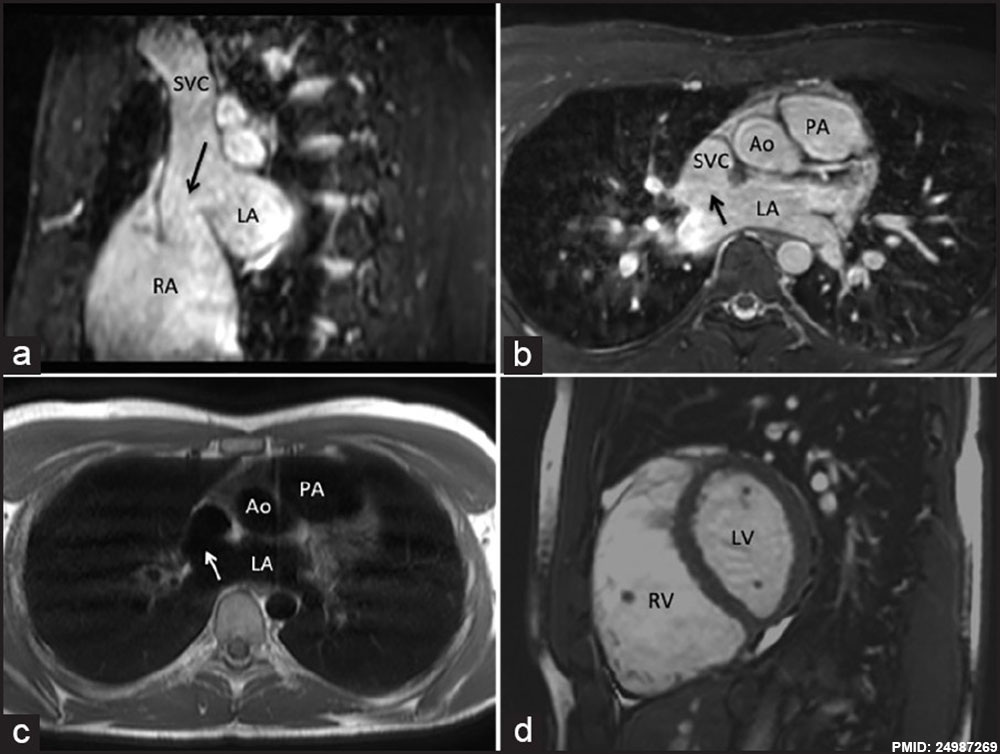
Figure 1: (a) Breath-held fat suppressed three-dimensional steady-state free precession (SSFP) pulse sequence in diastole in the sagittal view demonstrating sinus venosus atrial septal defect (SV-ASD) (arrow) between superior vena cava (SVC) and left atrium (LA). (b) Breath-held fat suppressed three-dimensional SSFP pulse sequence in diastole in the axial view demonstrating SV-ASD (arrow) between SVC and LA. (c) Turbo spin-echo black blood image in the same axial plane as ure 1b demonstrating SV-ASD (arrow) between SVC and LA. (d) SSFP image showing the dilated right ventricle (RV) and left ventricle (LV)
File history
Click on a date/time to view the file as it appeared at that time.
| Date/Time | Thumbnail | Dimensions | User | Comment | |
|---|---|---|---|---|---|
| current | 09:27, 22 February 2015 | | 1,000 × 754 (90 KB) | Z8600021 (talk | contribs) | http://www.annalspc.com/article.asp?issn=0974-2069;year=2014;volume=7;issue=2;spage=160;epage=162;aulast=Ganigara The role of cardiac MRI in the diagnosis and management of sinus venosus atrial septal defect. Ann Pediatr Cardiol. 2014 May;7(2):160-... |
You cannot overwrite this file.
File usage
There are no pages that use this file.