File:Keibel1910 fig28-29.jpg
Original file (1,700 × 520 pixels, file size: 51 KB, MIME type: image/jpeg)
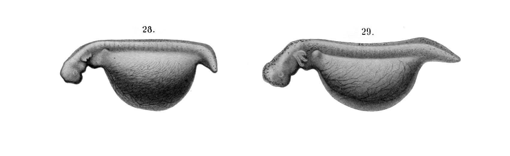
Note - Magnifications refer to original print versions.
Fig. 28. (X 5)
Side view of embryo 30 days 8 hrs. old, length 13 mm, 30 — 31 myotomes. The trunk of the embryo is nearly straight. At level of the posterior gill there is a pronounced neck bend and at the level of the posterior limbs a striking downward bend of the tail. The epiphysis shows in surface views. The lens is discernible. The ear is still vi.sible. The external nasal openings are sharply defined. The boundaries of the mouth are better outlined owing to the approximation of the ventral ends of the mandibular arches. The hyoid arch is becoming obscured. The gill bars are prominent on the three branchial arches. The anterior limb buds project dorsally about .5 mm above the surface of the bod3\ The posterior limb buds are but slight elevations. The yolk is pear-shaped with its dorsal surface much flattened. The auricular and ventricular portions of the heart are apparent. The surface of the yolk is covered by a dense network of capillaries which for the most part convey blood antero-ventrally to the abdominal vein. Considerable pigment is present in the trunk region although but little has reached the outer portion of the dermis.
Fig. 29. (X 5)
Side view of embryo 36 days 16 hrs. old, length 16 mm, 36-38 myotomes. In general outline the embryo shows a numljcr of striking changes. The neck bend is not so pronounced. The tail bend is scarcely noticeable. There is a striking increase in dorso-ventral width of tail. The cerebral hemispheres are well defined. The eye is now prominent and the lens better defined. The ear is no longer visible in surface views. The mouth is well defined. The ends of the mandibular arches are closely approximated but not united. The hyoid and branchial arches are more obscure. Anlagen of gill fiiaments present on gill bars. Anterior limbs project dorsally. Posterior limbs are short ridges extending in horizontal plane. The yolk is elongated and reduced in diameter both dorso-ventrally and laterally. Surface blood vessels as in preceding stage, excepting that they are now apparent in the gill bars. The chromatophores are most numerous in the anterior and dorsal portions of the head.
File history
Yi efo/eka'e gwa ebo wo le nyangagi wuncin ye kamina wunga tinya nan
| Gwalagizhi | Nyangagi | Dimensions | User | Comment | |
|---|---|---|---|---|---|
| current | 14:57, 10 January 2015 | 1,700 × 520 (51 KB) | Z8600021 (talk | contribs) | '''Note''' - Magnifications refer to original print versions. Fig. 28. (X 5) Side view of embryo 30 days 8 hrs. old, length 13 mm, 30 — 31 myotomes. The trunk of the embryo is nearly straight. At level of the posterior gill there is a pronounced n... |
You cannot overwrite this file.
File usage
The following 2 pages use this file: