File:Fetal pineal gland 01.jpg
From Embryology
Size of this preview: 696 × 600 pixels. Other resolution: 700 × 603 pixels.
Original file (700 × 603 pixels, file size: 67 KB, MIME type: image/jpeg)
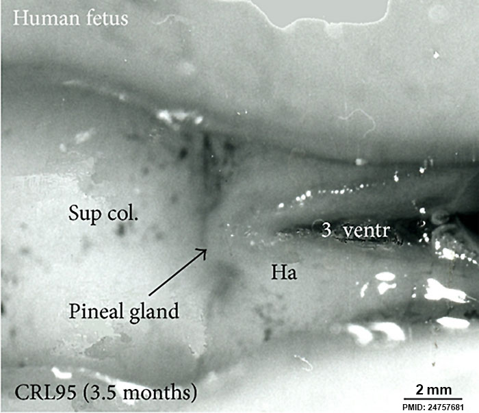
Superior (dorsal) view of the diencephalic-mesencephalic area of a 3.5-month-old human fetus. The third ventricle (3 ventr) without pial covering is seen to the right in the micrograph. The small pineal gland is a small protuberance (arrow) and merging via the broad stalk with the habenula (Ha). Sup col.: superior colliculus. Bar = 2 mm.
Figure 3: 868567.fig.003.jpg
File history
Click on a date/time to view the file as it appeared at that time.
| Date/Time | Thumbnail | Dimensions | User | Comment | |
|---|---|---|---|---|---|
| current | 09:05, 13 September 2014 | | 700 × 603 (67 KB) | Z8600021 (talk | contribs) | Superior (dorsal) view of the diencephalic-mesencephalic area of a 3.5-month-old human fetus. The third ventricle (3 ventr) without pial covering is seen to the right in the micrograph. The small pineal gland is a small protuberance (arrow) and mergi... |
You cannot overwrite this file.
File usage
The following 4 pages use this file: