File:Hilfer1990 Fig16.jpg
From Embryology
Size of this preview: 540 × 600 pixels. Other resolution: 1,200 × 1,333 pixels.
Original file (1,200 × 1,333 pixels, file size: 247 KB, MIME type: image/jpeg)
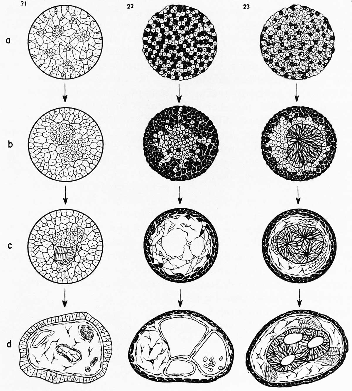
Figure 16. Illustration from Townes and Holtfreter (1955) demonstrating sorting of dissociated cells from different embryonic layers in amphibians. Cells from different germ layers sorted into clumps and tended to take a position resembling that of normal embryonic development.
File history
Click on a date/time to view the file as it appeared at that time.
| Date/Time | Thumbnail | Dimensions | User | Comment | |
|---|---|---|---|---|---|
| current | 10:42, 28 August 2014 | | 1,200 × 1,333 (247 KB) | Z8600021 (talk | contribs) | Figure 16. Illustration from Townes and Holtfreter (1955) demonstrating sorting of dissociated cells from different embryonic layers in amphibians. Cells from different germ layers sorted into clumps and tended to take a position resembling that of nor... |
You cannot overwrite this file.
File usage
The following page uses this file: