File:Hilfer1990 Fig05.jpg

Original file (1,096 × 1,200 pixels, file size: 93 KB, MIME type: image/jpeg)
Figure 5.
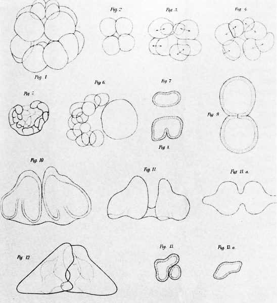
Illustrations from H. Driesch (1892) showing effects of manipulating blastomeres of sea urchin eggs. His Figure 1 shows a normal 16-cell stage (copied from Selenka). Figure 2 shows a half-embryo from an 8-cell stage and Figures 3 and 4 half-embryos from a 16-cell stage. Figure 5 is a half-embryo at the blastula stage. These half-embryos developed into complete individuals. Figures 7 through 9 show blastulae in the process of dividing. These formed conjoined twins as shown in Figures 10 through 12. These results were interpreted as demonstrating regulative development.
(Courtesy of the Marine Biological Laboratory.)
File history
Yi efo/eka'e gwa ebo wo le nyangagi wuncin ye kamina wunga tinya nan
| Gwalagizhi | Nyangagi | Dimensions | User | Comment | |
|---|---|---|---|---|---|
| current | 09:26, 28 August 2014 |  | 1,096 × 1,200 (93 KB) | Z8600021 (talk | contribs) | ==Figure 5. == Illustrations from H. Driesch (1892) showing effects of manipulating blastomeres of sea urchin eggs. His Figure 1 shows a normal 16-cell stage (copied from Selenka). Figure 2 shows a half-embryo from an 8-cell stage and Figures 3 and 4... |
You cannot overwrite this file.
File usage
The following 3 pages use this file: