File:Mouse Brain E17 MRI 01.jpg
Original file (963 × 403 pixels, file size: 88 KB, MIME type: image/jpeg)
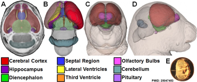
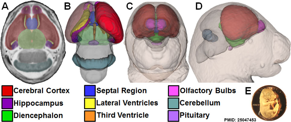
Magnetic resonance microscopy (MRM) enables concurrent visualization of the brain and face of GD17 mouse fetuses. Forebrain regions, pituitary, and cerebellum were manually segmented from transverse 39 µm MRM sections (A). 3D brain reconstructions were generated by overlaying manually segmented regions with whole-brain masks (B). Reduced opacity of the left cortex and diencephalon allows visualization of the left ventricle, hippocampus, third ventricle, and pituitary. From the same MRM scans, 3D head reconstructions were created, allowing concurrent visualization of the face and brain in situ (C–D). The size of a GD17 mouse fetus can be appreciated when shown in scale with a U.S. penny (E).
Figure 1. doi:10.1371/journal.pone.0102603.g001
File history
Yi efo/eka'e gwa ebo wo le nyangagi wuncin ye kamina wunga tinya nan
| Gwalagizhi | Nyangagi | Dimensions | User | Comment | |
|---|---|---|---|---|---|
| current | 19:02, 27 August 2014 | | 963 × 403 (88 KB) | Z8600021 (talk | contribs) | Magnetic resonance microscopy (MRM) enables concurrent visualization of the brain and face of GD17 mouse fetuses. Forebrain regions, pituitary, and cerebellum were manually segmented from transverse 39 µm MRM sections (A). 3D brain reconstructions wer... |
You cannot overwrite this file.
File usage
The following page uses this file: