File:Gray0463.jpg
Original file (1,099 × 755 pixels, file size: 134 KB, MIME type: image/jpeg)
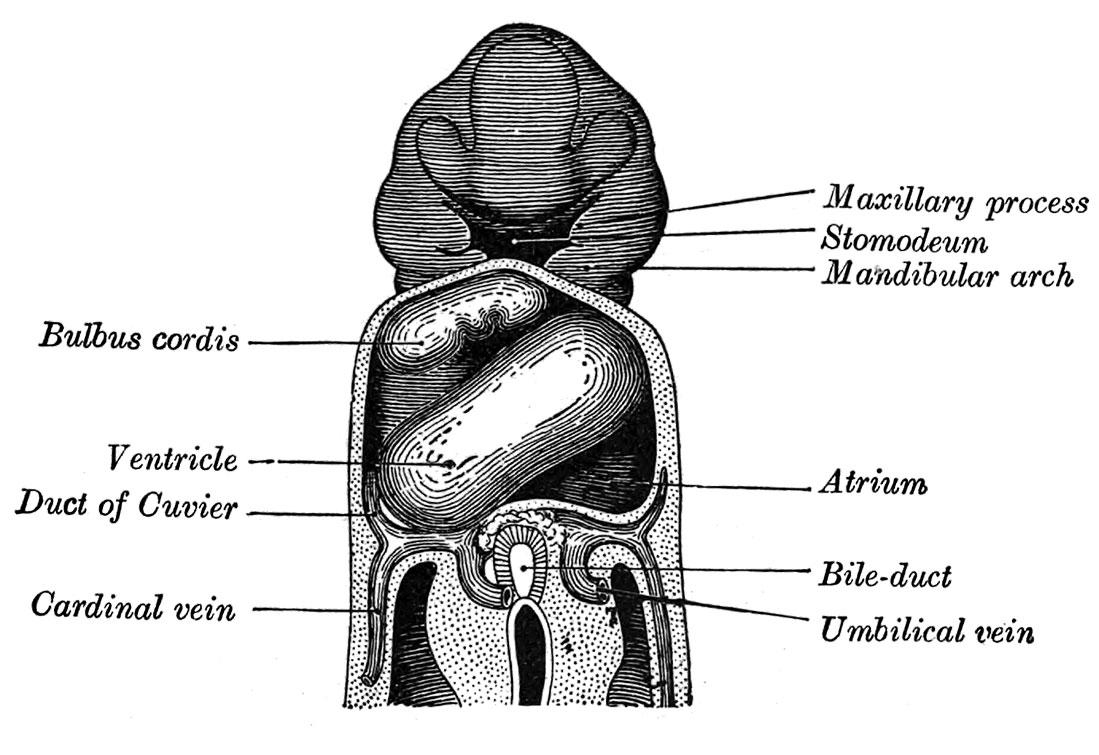
Fig. 463. Heart of human embryo of about fifteen days
Reconstruction by Wilhelm His (1831-1904)
The sinus venosus is at first situated in the septum transversum (a layer of mesoderm in which the liver and the central tendon of the diaphragm are developed) behind the primitive atrium, and is formed by the union of the vitelline veins. The veins or ducts of Cuvier from the body of the embryo and the umbilical veins from the placenta subsequently open into it (Fig. 463) . The sinus is at first place transversely, and opens by a median aperture into the primitive atrium.
| Historic Disclaimer - information about historic embryology pages |
|---|
 |
- Gray's Images: Development | Lymphatic | Neural | Vision | Hearing | Somatosensory | Integumentary | Respiratory | Gastrointestinal | Urogenital | Endocrine | Surface Anatomy | iBook | Historic Disclaimer
| Historic Disclaimer - information about historic embryology pages |
|---|
|
| iBook - Gray's Embryology | |
|---|---|

|
|
Reference
Gray H. Anatomy of the human body. (1918) Philadelphia: Lea & Febiger.
Cite this page: Hill, M.A. (2024, June 15) Embryology Gray0463.jpg. Retrieved from https://embryology.med.unsw.edu.au/embryology/index.php/File:Gray0463.jpg
- © Dr Mark Hill 2024, UNSW Embryology ISBN: 978 0 7334 2609 4 - UNSW CRICOS Provider Code No. 00098G
File history
Click on a date/time to view the file as it appeared at that time.
| Date/Time | Thumbnail | Dimensions | User | Comment | |
|---|---|---|---|---|---|
| current | 18:47, 23 August 2014 | | 1,099 × 755 (134 KB) | Z8600021 (talk | contribs) | |
| 18:47, 23 August 2014 |  | 1,200 × 814 (149 KB) | Z8600021 (talk | contribs) |
You cannot overwrite this file.
File usage
The following page uses this file: