File:Dog patent ductus arteriosus computed tomography.jpg
Original file (600 × 654 pixels, file size: 101 KB, MIME type: image/jpeg)
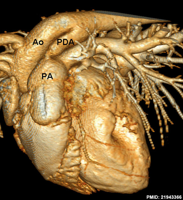
Dog Patent Ductus Arteriosus Computed Tomography
Volume-rendered 3-dimensional multidetector-row computed tomography (MDCT) reconstruction of a patent ductus arteriosus. The patent ductus arteriosus (PDA) connects the descending aorta dorsally to the main pulmonary artery.
- Ao - aorta
- PA - pulmonary artery
- PDA - patent ductus arteriosus
- Links: Patent Ductus Arteriosus | Cardiovascular System Development | Computed Tomography | Dog Development
Reference
<pubmed>21943366</pubmed>| BMC Vet Res.
Copyright
© 2011 Henjes et al; licensee BioMed Central Ltd. This is an Open Access article distributed under the terms of the Creative Commons Attribution License (http://creativecommons.org/licenses/by/2.0), which permits unrestricted use, distribution, and reproduction in any medium, provided the original work is properly cited.
Figure 2. Modified in size and labelling.
File history
Click on a date/time to view the file as it appeared at that time.
| Date/Time | Thumbnail | Dimensions | User | Comment | |
|---|---|---|---|---|---|
| current | 22:58, 25 June 2014 | | 600 × 654 (101 KB) | Z8600021 (talk | contribs) | ==Dog Patent Ductus Arteriosus Computed Tomography== ===Reference=== <pubmed>21943366</pubmed>| [http://www.biomedcentral.com/1746-6148/7/57 BMC Vet Res.] ====Copyright==== © 2011 Henjes et al; licensee BioMed Central Ltd. This is an Open Access... |
You cannot overwrite this file.
File usage
The following page uses this file: