File:Human oocyte em01.jpg

Human_oocyte_em01.jpg (600 × 589 pixels, file size: 65 KB, MIME type: image/jpeg)
Human Oocyte Cortical Granules and Microvilli (EM)
Cortical granules (CG) and microvilli (MV) in oocytes from control and oocytes with a dark zona pellucid. A rim of electron-dense cortical granules (arrows) was observed just beneath the oolemma of the MII oocytes with a DZP and MII oocytes in the control group (B, A). Microvilli are numerous and long on the oolemma in each group (A, B). MV: microvilli; CG: cortical granules; PVS: perivitelline space; O: oocyte. Scale bar (A, B): 500 nm.
Reference
<pubmed>24586757</pubmed>| PMC3933533 | PLoS One
Copyright
© 2014 Shi et al. This is an open-access article distributed under the terms of the Creative Commons Attribution License, which permits unrestricted use, distribution, and reproduction in any medium, provided the original author and source are credited.
doi:10.1371/journal.pone.0089409.g004
Original image panel cropped from figure 4, resized, recoloured and relabelled.
Journal.pone.0089409.g004.jpg
File history
Click on a date/time to view the file as it appeared at that time.
| Date/Time | Thumbnail | Dimensions | User | Comment | |
|---|---|---|---|---|---|
| current | 11:39, 24 June 2014 | | 600 × 589 (65 KB) | Z8600021 (talk | contribs) | |
| 12:09, 19 June 2014 | 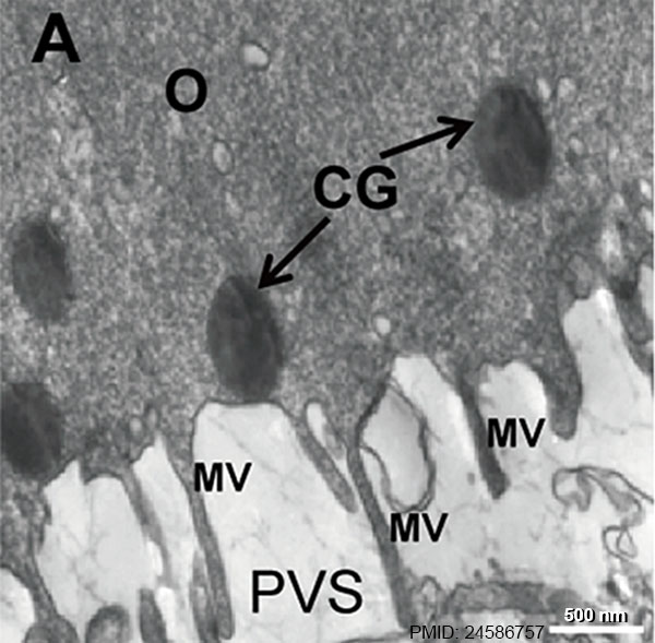 | 600 × 589 (65 KB) | Z8600021 (talk | contribs) | ==Human Oocyte Cortical Granules and Microvilli (EM)== Cortical granules (CG) and microvilli (MV) in oocytes from control and oocytes with a dark zona pellucid. A rim of electron-dense cortical granules (arrows) was observed just beneath the oolemma o... |
You cannot overwrite this file.
File usage
There are no pages that use this file.