File:Mouse oocyte balbini body EM01.jpg
From Embryology

Size of this preview: 595 × 599 pixels. Other resolution: 695 × 700 pixels.
Original file (695 × 700 pixels, file size: 155 KB, MIME type: image/jpeg)
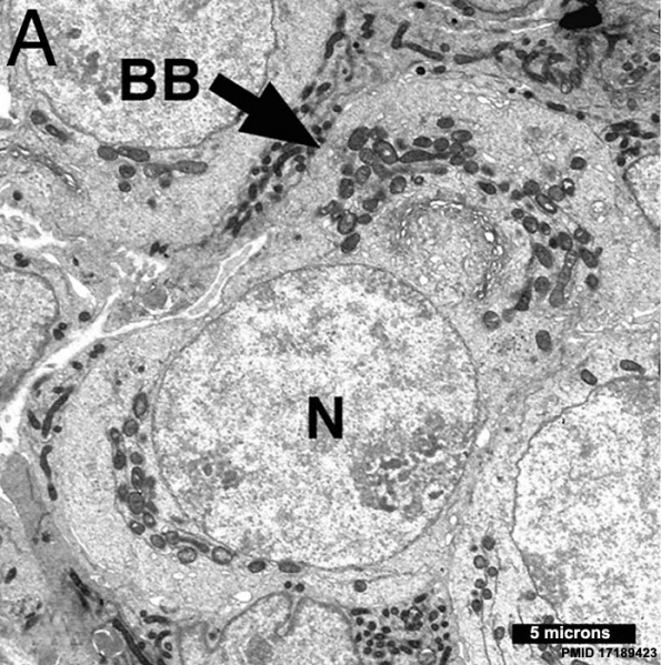
Mouse Oocyte Balbini Body
Electron micrograph of oocytes in neonatal ovary micrograph of an oocyte within a germline cyst from PND1 showing a well defined Balbiani body (arrow) with Golgi surrounded by mitochondria.
- Balbiani body (mitochondrial cloud) is a large organelle aggregate found in developing oocytes of many species.
- EM Links: All Images | balbini_body | A | B | C | D | E | F | Oocyte Development | Ovary Development | Mouse Development
Reference
<pubmed>17189423</pubmed>| PMC1765432 | Proc Natl Acad Sci U S A.
Copyright
Proceedings National Academy of Sciences (PNAS) Liberalization of PNAS copyright policy: Noncommercial use freely allowed Note original Author should be contacted for permission to reuse for Educational purposes. See also PNAS Author Rights and Permission FAQs
- Cozzarelli NR, Fulton KR, Sullenberger DM. Liberalization of PNAS copyright policy: noncommercial use freely allowed. Proc Natl Acad Sci U S A. 2004 Aug 24;101(34):12399. PMID15314225 "Our guiding principle is that, while PNAS retains copyright, anyone can make noncommercial use of work in PNAS without asking our permission, provided that the original source is cited."
File history
Yi efo/eka'e gwa ebo wo le nyangagi wuncin ye kamina wunga tinya nan
| Gwalagizhi | Nyangagi | Dimensions | User | Comment | |
|---|---|---|---|---|---|
| current | 13:43, 9 May 2013 | | 695 × 700 (155 KB) | Z8600021 (talk | contribs) | ==Mouse oocyte balbini body== Electron micrographs of oocytes in neonatal ovaries. (A) Micrograph of an oocyte within a germline cyst from PND1 showing a well defined Balbiani body (arrow) with Golgi surrounded by mitochondria. {{PNAS}} |
You cannot overwrite this file.
File usage
The following 2 pages use this file: