File:Placental chorioangioma ultrasound 01.jpg
Original file (1,183 × 837 pixels, file size: 101 KB, MIME type: image/jpeg)
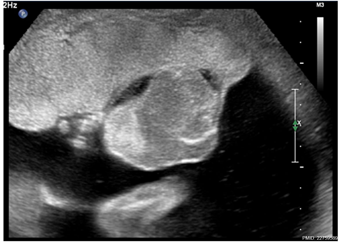
Placental Chorioangioma Ultrasound
Gross examination of the placenta revealed a yellowish, well-circumscribed firm mass measuring 5 cm × 5 cm connected by two vessels to the placenta.
Histopathologic examination revealed a placental disc 15 cm × 17 cm × 13 cm, with a three-vessel umbilical cord that was attached peripherally and measured 9 cm × 1.5 cm. The weight of the placenta was 530 g. The tumor was confirmed to be a chorioangioma.
- Links: Ultrasound scan | Ultrasound blood flow | Gross image 1 | Gross image 2 | Gross image 3 | Placental Chorioangioma | Placenta - Abnormalities
Reference
<pubmed>22759589</pubmed>| PMC3419096 | J Med Case Rep
Copyright
© 2012 Babic et al.; licensee BioMed Central Ltd. This is an Open Access article distributed under the terms of the Creative Commons Attribution License (http://creativecommons.org/licenses/by/2.0), which permits unrestricted use, distribution, and reproduction in any medium, provided the original work is properly cited.
Figure 5. Original image was adjusted in size, contrast and labelling.
1752-1947-6-183-1.jpg
File history
Yi efo/eka'e gwa ebo wo le nyangagi wuncin ye kamina wunga tinya nan
| Gwalagizhi | Nyangagi | Dimensions | User | Comment | |
|---|---|---|---|---|---|
| current | 11:19, 7 June 2014 | | 1,183 × 837 (101 KB) | Z8600021 (talk | contribs) | 1752-1947-6-183-1.jpg |
You cannot overwrite this file.
File usage
The following page uses this file: