File:Mouse oocyte Egr3-01.jpg
From Embryology
Size of this preview: 537 × 600 pixels. Other resolution: 600 × 670 pixels.
Original file (600 × 670 pixels, file size: 152 KB, MIME type: image/jpeg)
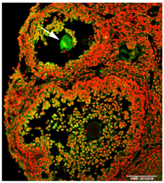
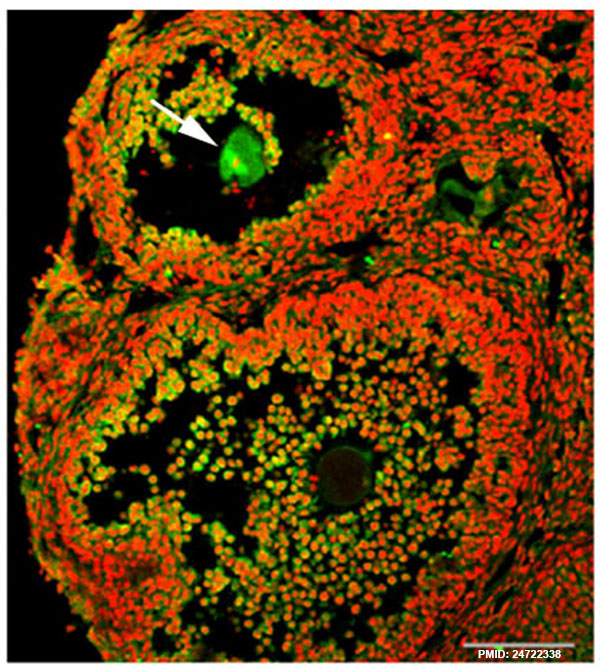
While investigating the expression of Egr transcription factors in the mouse ovary, we unexpectedly observed that Egr3 was localized to the meiotic spindles of maturing oocytes (Fig. 1A, arrow).
Journal.pone.0094708.g001.jpg
File history
Yi efo/eka'e gwa ebo wo le nyangagi wuncin ye kamina wunga tinya nan
| Gwalagizhi | Nyangagi | Dimensions | User | Comment | |
|---|---|---|---|---|---|
| current | 22:38, 16 April 2014 | | 600 × 670 (152 KB) | Z8600021 (talk | contribs) | While investigating the expression of Egr transcription factors in the mouse ovary, we unexpectedly observed that Egr3 was localized to the meiotic spindles of maturing oocytes (Fig. 1A, arrow). Journal.pone.0094708.g001.jpg |
You cannot overwrite this file.
File usage
There are no pages that use this file.