File:Minot1889 fig26.jpg
From Embryology
Size of this preview: 627 × 599 pixels. Other resolution: 952 × 910 pixels.
Original file (952 × 910 pixels, file size: 336 KB, MIME type: image/jpeg)
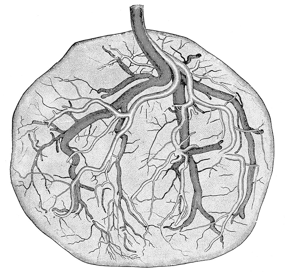
Cut 26. Placenta at full term, doubly injected
by Df. H. P. Quincy, to show the distribution of blood-vessels upon the surface; the arteries are drawn light; the veins dark. X 0.7 diams.
Reference
Minot CS. Uterus And Embryo - I. Rabbit II. Man. (1889) J Morphol. 2:
Cite this page: Hill, M.A. (2024, June 26) Embryology Minot1889 fig26.jpg. Retrieved from https://embryology.med.unsw.edu.au/embryology/index.php/File:Minot1889_fig26.jpg
- © Dr Mark Hill 2024, UNSW Embryology ISBN: 978 0 7334 2609 4 - UNSW CRICOS Provider Code No. 00098G
File history
Yi efo/eka'e gwa ebo wo le nyangagi wuncin ye kamina wunga tinya nan
| Gwalagizhi | Nyangagi | Dimensions | User | Comment | |
|---|---|---|---|---|---|
| current | 12:57, 4 April 2014 | | 952 × 910 (336 KB) | Z8600021 (talk | contribs) | no legend |
| 12:57, 4 April 2014 |  | 952 × 1,000 (357 KB) | Z8600021 (talk | contribs) | {{Minot1889 figures}} |
You cannot overwrite this file.
File usage
The following page uses this file: