File:Human embryo neck 01.jpg

Original file (534 × 827 pixels, file size: 186 KB, MIME type: image/jpeg)
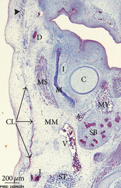
Human Embryo Neck Region
Human embryo GI4 (26.5 mm GL; 8 weeks of development). Frontal section. Azocarmine staining. The cervical lamina (CL) extends towards the mandibular region enclosing the masseter muscle (MS). Arrowhead: infraorbital lamina.
Bar: 200 μm.
- A - Facial artery
- C - Meckel’s cartilage
- CL - Cervical lamina
- D - Parotid duct
- I - Inferior alveolar nerve
- J - External jugular vein
- MM - Marginal mandibular branch of the facial nerve
- MS - Masseter muscle
- MY - Mylohyoid muscle
- SB - Submandibular gland
- ST - Sternocleidomastoid muscle
- V - Facial vein
- Human Neck Links: Week 8 Frontal section overview | Week 8 Frontal section detail | Week 8 Parotid region | Week 10 Frontal section | Neck muscle cartoon
Reference
<pubmed>24396304 </pubmed>| ScientificWorldJournal.
Copyright
Copyright © 2013 C. De la Cuadra-Blanco et al. This is an open access article distributed under the Creative Commons Attribution License, which permits unrestricted use, distribution, and reproduction in any medium, provided the original work is properly cited.
Figure 1: (a) 716962.fig.001a.jpg
File history
Yi efo/eka'e gwa ebo wo le nyangagi wuncin ye kamina wunga tinya nan
| Gwalagizhi | Nyangagi | Dimensions | User | Comment | |
|---|---|---|---|---|---|
| current | 11:46, 28 March 2014 | | 534 × 827 (186 KB) | Z8600021 (talk | contribs) | ==Human Embryo Neck Region== Human embryo GI4 (26.5 mm GL; 8 weeks of development). Frontal section. Azocarmine staining. The cervical lamina (CL) extends towards the mandibular region enclosing the masseter muscle (MS). Arrowhead: infraorbital lami... |
You cannot overwrite this file.
File usage
There are no pages that use this file.