File:Placenta percreta 02.jpg
Original file (1,000 × 405 pixels, file size: 105 KB, MIME type: image/jpeg)
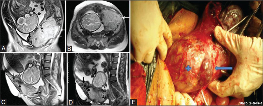
Figure 10 (A-E): Low-lying anterior placenta percreta in a 32-year-old woman at 34 weeks of gestation. (A, B) Coronal and axial T2 HASTE MR images show a heterogeneous placenta with thick, dark intraplacental bands (black arrows). Focal interruption of the left inferolateral myometrium and subserosal extension of placental tissue (white arrows). (C, D) Sagittal T2 HASTE and T2 fast spin echo (FSE) MR images show focal interruption of anterior myometrium (black arrowheads), anterosuperior to the internal os and extending subserosally (white arrowheads). Marked heterogeneity of the anterior myometrium, just superior to the invasion noted, represents abnormal vascularity (arrow). (E) Surgical photograph showing the placenta extending through uterine wall (+) and covered by thin serosal layer (arrow). No features of bladder invasion
File history
Yi efo/eka'e gwa ebo wo le nyangagi wuncin ye kamina wunga tinya nan
| Gwalagizhi | Nyangagi | Dimensions | User | Comment | |
|---|---|---|---|---|---|
| current | 09:59, 19 March 2014 | | 1,000 × 405 (105 KB) | Z8600021 (talk | contribs) | Figure 10 (A-E): Low-lying anterior placenta percreta in a 32-year-old woman at 34 weeks of gestation. (A, B) Coronal and axial T2 HASTE MR images show a heterogeneous placenta with thick, dark intraplacental bands (black arrows). Focal interruption of... |
You cannot overwrite this file.
File usage
There are no pages that use this file.


